https://www.ncbi.nlm.nih.gov/pubmed/7495135
(email me if you want a copy of the article)
- Upper respiratory tract
- Fallopian tube and parts of the endometrium
- Ependymal cells that line the ventricles in the brain
- Caput epididymis/efferent ducts
- Any number of other locations where the epithelium has undergone a change called ciliary metaplasia (the epithelium converts from non-ciliated cells to ciliated cells)
This was definitely a challenging case, and I loved seeing the great discussion that it generated! Huge kudos to Michigan Micro who pointed out the diagnostic features and led everyone away from a diagnosis of Trichomonas vaginalis (which was the predominant answer for quite a while). These key features are:
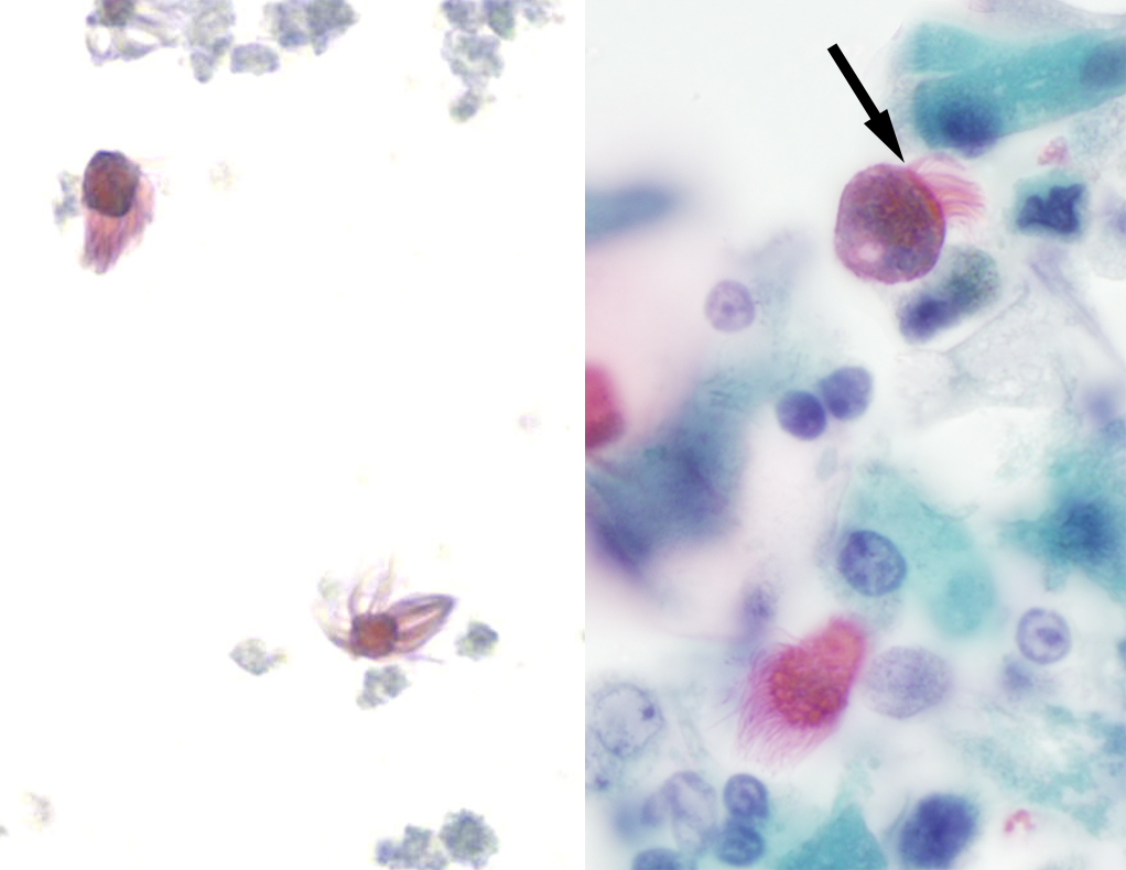
Reason #1. T. vaginalis doesn't have as many flagella as are seen in this case, and the flagella are more spaced out rather than grouped at one end. Compare the flagella of Trichomonas (top photo below, arrow) to the dense mat of cilia (arrow) on respiratory epithelial cells (bottom image):

The bottom right image looks a lot like the following still photos taken from this case:

Reason #2: T. vaginalis has an undulating membrane, which is absent here (although it is hard to tell from the video).
Reason #3: T. vaginalis has 'jerky' motility (see my previous case of the week for trichomonad motility HERE - warning for obnoxious music)
Reason #4: T. vaginalis is usually more rounded or pear-shaped, whereas this specimen is more elongated.
The last component to consider is size, which unfortunately was not provided with the case. However, here are sizes of similar-appearing objects for comparison:
- Trichomonas vaginalis trophozoites: 7-30 microns x 6-15 microns
- Detached ciliary tufts: 10-15 microns diameter
- Intact ciliated columnar epithelial cells: Generally 4 times longer than wide, so this would be approximately 40-60 microns long x 10-15 microns wide.
- Balantidium coli trophozoites: 40-100 microns in greatest dimension


3 comments:
It is a very interesting case and every week we learn something new in this blog. Thank you Dr. Pritt for sharing it and thank for Michigan Micro who pointed out that it's not what we have thought.
The only reason I knew it was a ciliated cell was from reading this blog every week. So, thank Dr. Pritt for sharing her knowledge. This blog is one of the best parasitology resources around.
Michigan Micro
Thank you for the kind comments Yasir and Michigan Micro! I really appreciate the feedback and am glad that you find the blog to be helpful.
Bobbi
Post a Comment