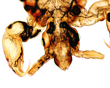
Thanks to Idzi for another spectacular case. This one nicely compliments his last case (see Case of the Week 509) which features a different round worm that can also be found in the eye: Dirofilaria. I've seen several ocular dirofilariasis cases in the past few years - all which had been initially mistaken for Loa loa - so I think it's important for microbiologists and clinicians to know that the two worms can have a similar presentation.
So how can you tell them apart? Here are some helpful features:
1. Travel history: Loa loa is found in West and Central Africa, whereas Dirofilaria has a much broader distribution (Africa, Asia, Europe). If the patient hasn't been to West or Central Africa, then the diagnosis is probably not loiasis.
2. Morphology of the adult worm: The adults of both worms have a similar size and gross appearance. However, Loa loa has irregularly-spaced cuticular bosses ('bumps' on the outer aspect of the cuticle), whereas Dirofilaria has longitudinal ridges. Here are some representative images of the two:
Loa loa (I sadly neglected to include this image from the case earlier):
Dirofilaria repens:
3. Morphology of the microfilariae: Last, but not least, the morphology of the blood microfilariae is another helpful feature for differentiating L. loa from Dirofilaria. While the former are sheathed and have nuclei that go to the tip of the tail, the latter are rarely seen in human blood, are NOT sheathed, and the nuclei do NOT go to the tip of the tail.
Loa loa:
Dirofilaria repens:Note that the sheath of Loa loa (as well as Wuchereria bancrofti and Brugia timori) is usually colorless with tradition Giemsa stain, but is highlighted using Carazzi's hematoxylin stain as shown here. In comparison, the sheath of Brugia malayi is usually - but not always! - bright pink on Giemsa stain.


No comments:
Post a Comment