Wednesday, October 31, 2018
Halloween Parasite #3
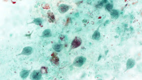
Happy Halloween Week everyone! As you all know, parasites can be creepy dreadful, but also fascinating, and sometimes even helpful. As a special Halloween treat, I'll be highlighting 5 different parasites on the Mayo Clinic News Network - 1 each day. HERE is parasite #3 - . These are written as educational pieces for the general public. Feel free to use the text and images for your own educational purposes.
Tuesday, October 30, 2018
Halloween Case #2
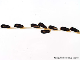
Happy Halloween Week everyone! As you all know, parasites can be creepy dreadful, but also fascinating, and sometimes even helpful. As a special Halloween treat, I'll be highlighting 5 different parasites on the Mayo Clinic News Network - 1 each day. HERE is parasite #2 - scalp explorers. These are written as educational pieces for the general public. Feel free to use the text and images for your own educational purposes.
Monday, October 29, 2018
Halloween Case #1
Happy Halloween Week everyone! As you all know, parasites can be creepy dreadful, but also fascinating, and sometimes even helpful. As a special Halloween treat, I'll be highlighting 5 different parasites on the Mayo Clinic News Network HERE - 1 each day.
I unfortunately will not be having a Halloween party this year since I am at the annual meeting of the American Society of Tropical Medicine and Hygiene in New Orleans. Therefore I won't have any costume photos from my party, but I promise to post photos later if I see any particularly parasite-like costumes on Bourbon street!
I unfortunately will not be having a Halloween party this year since I am at the annual meeting of the American Society of Tropical Medicine and Hygiene in New Orleans. Therefore I won't have any costume photos from my party, but I promise to post photos later if I see any particularly parasite-like costumes on Bourbon street!
Monday, October 22, 2018
Case of the Week 516
This object was removed and placed in a sterile container without preservative:
It was still quite lively!
Approximately 1 week later, it was sent to my lab for identification. Even though it had darkened significantly, the diagnostic features were still apparent. Here is the posterior end:
Identification?
Sunday, October 21, 2018
Answer to Case 516
Answer: Cordylobia anthropophaga, the mango or tumbu fly.
As Florida Fan nicely outlined, there are several initial features that lead us to the identification:
1. The patient is from Africa, where we can find the tumbu fly.
2. The size of the larva is about one third or one fourth the diameter of the cup, roughly 13-15 mm in length, compatible to that of this fly's larva size.
3. There is no discernible peritreme nor ecdysial scar.
4. The spiracles open through sinuous slits.
The following image shows the posterior spiracles, with the sinuous slits (arrow head). Note that the peritreme is not easily discernible (arrow).
Blaine and I covered this classic causes of myiasis in our CAP Arthropod Benchtop Reference Guide. The key morphologic features are its robust form, cuticular spines on all body segments (not clearly shown here), spiracular plat with a very weak peritreme, and sinuous slits. As Blaine pointed out, you can differentiate C. anthropophaga from the similar-appearing species, C. rodhaini, by its much more sinuous slits.
Monday, October 15, 2018
Case of the Week 515
This week's case is a "worm" found in the cecum - an incidental finding during colonoscopy. It measures approximately 4 cm long. Identification? Images by my Parasitology technologist extraordinaire, Heather Rose Arguello.
Sunday, October 14, 2018
Answer to Case 515
Answer: Trichuris trichiura, a.k.a. "whipworm"
As noted by Florida Fan, "we can clearly see the thread-like anterior portion and an inflated posterior, giving the worm resemblance to a whip". I always like to ask my students which end they think is anterior - they invariably (incorrectly) guess that it's the broader end! This gives me the opportunity to walk them through the life cycle and how the adult worms are embedded within the mucosa of the large bowel. It always seems to make more sense to them when I ask which end would be easier to thread into the wall of the bowel - the thin end or the thick end? This helps explain why the anterior end is thinner than the posterior. I also note that having a broader posterior end allows it to fill with eggs in the female worm, and these can then easily be shed into the intestinal lumen . I'm kind of old-fashioned, so my explanation is usually accompanied by some hand-drawn images.
Florida Fan also noted another two other important morphologic features: the stichosome comprised of multiple stichocytes, and the immature ova within the uterus in the posterior end. The purpose of the stichosome is unclear, but it appears to serve a secretory function. It is found posterior to the esophagus and is quite prominent in some nematodes such as Trichuris species.
Check out the comments for a poem by Blaine referencing the dreaded complication of whipworm infection - rectal prolapse!
Monday, October 8, 2018
Case of the Week 514
This week's case is a classic case from Heather Rose Arguello, my fabulous parasitology specialist. This is a wet preparation of liver cyst fluid (40x objective):
Identification?
Identification?
Sunday, October 7, 2018
Answer to Case 514
Answer: Hydatid sand of Echinococcus sp.; mostly likely Echinococcus granulosus due to the abundant protoscoleces/hooklets and presence within a single cyst.
The photo from this case shows numerous free hooklets in the background of degenerating protoscoloces. As Blaine pointed out, the mixture of disintegrating protoscoleces with free hooklets and calcareous corpuscles is often referred to as "hydatid sand."
As Old One pointed out, the protoscoleces have a rostellum with 2 rows of hooklets - one row of large hooklets and one row of small hooklets - making this cestode "armed". He further notes that having an armed rostellum puts this cestode into the family Taeniidae.
Echinococcus species are one of several members of the Taeniidae that can cause human disease. Echinococcus granulosus is now recognized as a complex of closely-related organisms, with E. granulosus sensu stricto being the most common species causing human disease worldwide. Other members of this complex that cause human disease are E. canadensis and E. ortleppi. Differentiation of the members of the E. granulosus complex is primarily accomplished through molecular means.
E. multilocularis human infection is fortunately less common, as this parasite is known for its more aggressive and invasive growth pattern. As several readers pointed out, the cysts of E. multilocularis invade host tissue, much like a tumor, and are not contained within a large parent cyst. Protoscoleces are not commonly seen in human infections with E. multilocularis. Finally, E. oligarthra and E. vogeli are rare causes of human echinococcosis in South and Central America.
Monday, October 1, 2018
Case of the Week 513
This week features our monthly case from Idzi Potters and the Institute of Tropical Medicine, Antwerp. A Nigerian male patient, returns to Belgium, after having visited his relatives. He presents at the hospital, where a worm was extracted from his eye (length: 5 cm).
In a drop of blood, live “larvae” were seen.
A KNOTT concentration was made and stained with Carazzi’s to visualize the details.
The “larvae” are measuring (on average) 280 µm in length.
Diagnosis please?
In a drop of blood, live “larvae” were seen.
The “larvae” are measuring (on average) 280 µm in length.
Diagnosis please?


