See them in action!
Monday, February 25, 2019
Case of the Week 533
This week's case is from the very cool Belize Vector & Ecology Center run by Drs. Nicole L. Achee and John P. Grieco in Orange Walk, Belize. These arthropods are being reared to better understand their role as vectors of a human pathogen in this region. What pathogen is this?
Sunday, February 24, 2019
Answer to Case 533
Answer to Case of the Week 533:
The vector: Triatomine bugs (also called reduviid bugs, “kissing” bugs, and cone-nosed bugs).
The pathogen: Trypanosoma cruzi
Resultant disease: Chagas disease, a.k.a. American trypanosomiasis
Thank you all for the great comments! Hopefully we will see more cases from the Achee/Grieco lab in the future.
Monday, February 18, 2019
Case of the Week 532
This week's case was generously donated by Dr. Richard Bradbury and Dr. Sarah Sapp from the Morphology lab at the US Centers for Disease Control and Prevention. The following objects were seen in a wet mount of a concentrated stool specimen. The specimen was obtained from a West African baboon, but this finding may also be seen in human stool specimens. Identification?
Sunday, February 17, 2019
Answer to Case 532
Answer to Parasite Case of the Week 532: cyst and trophozoite of Balantioides, (a.k.a. Neobalantidium, formerly Balantidium) coli
This cool large ciliate is one of my favorite parasites. It's the largest protozoan parasite and only pathogenic ciliate to infect humans. As my predecessor, Dr. John H. Thompson, used to say, it is the aircraft carrier of the fecal flotilla.
Diagnosis is made based on the characteristic morphologic features. As Florida Fan mentioned, "At first look, the spherical object on the left of the first picture and the one of the third picture may lure us to identify them as eggs of some sort," (indeed, B. coli cysts may be easily mistaken for helminth eggs), "but the oval large object on the right side of the first picture as well as the second photo when enlarged will show us the cilia. The second picture shows also a cytostome (oral groove) on the upper end of the object."
Here is a closer look at these morphologic features of the trophozoite:
The 'kidney bean' shaped macronucleus is, unfortunately, not visible in these photos. It is another helpful diagnostic feature.
So what's up with the taxonomy? Well Blaine Mathison and I just published an update on parasite taxonomy discussing the new name, Neobalantidium, so imagine my dismay when I realized that the older (and potentially valid name) of Balantioides had already been described. As Blaine mentioned in his comment "It looks like when Neobalantidium was described in 2013, the authors overlooked a paper from 1931 that proposed the name Balantioides. If it was simply an oversight on their part, then yes the name could indeed be Balantioides. However, it is possible there was something in the description of Balantioides that rendered the name invalid (i.e., rules of nomenclature where not followed, at least for what was acceptable at the time). There is also another nomenclatural rule that states if a name is not used in the literature for a specific amount of time, it can be rendered invalid (not sure yet it that applies here)." Perhaps some of my readers can provide more insight. We are also checking with our colleagues to get more information and will report back to you, dear readers, when we have more answers.
Monday, February 11, 2019
Case of the Week 531
This week's case was generously donated by Florida Fan. The following was noted in skin scrapings from an elderly man.
This moving object was still noted 5 days after collection!
Identification?
This moving object was still noted 5 days after collection!
Identification?
Sunday, February 10, 2019
Answer to Case 531
Answer to Parasite Case of the Week 531: Sarcoptes scabei, var. hominis, the human "itch" mite. Seen here are mites, eggs and fecal pellets (scybala).
Old one nicely described the biology and morphology of these arthropods:
Mite shown in cross-section within the stratum corneum in a hematoxylin and eosin (H&E) stained tissue:
As noted by Bernardino Rocha, the scabies mite is an efficient miner. That's a neat way to think about this interesting arthropod. Sarcoptes scabei spend their entire lifecycle on or in their human host. The female burrows into the outer most layer of the epidermis (the stratum corneum) to create a short burrow called a molting pouch. The male penetrates this pouch and mates with the female. Interestingly, a female only mates once, and then remains fertile for the rest of her life. She leaves the molting pouch after mating to find another suitable location to create a permanent burrow. Bernardino further notes that "dispersed along the burrows are eggs, hatched larvae, and excrements."
According to the CDC, "Scabies mites generally do not survive more than 2 to 3 days away from human skin." However, the mite in this case was still 'alive and kicking' 5 days after collection, so clearly there is a range in the survival time away from the host, which may have important implications for scabies prevention and control measures.
I'll leave you all with this lovely poem from Blaine Mathison:
Old one nicely described the biology and morphology of these arthropods:
Sarcoptes scabei occur in a number of host species. Primarily in swine here in Minnesota but occasionally in humans. The male mites range in size from 213-285 μm long by 162-240 μm wide and the female mites range from 300-504 μm long to 230-420 μm wide. Sarcoptes are round to ovoid when viewed from the back; when viewed from the side they are ventrally flattened and dorsally rounded (similar to a turtle). They possess short stumpy legs, and have no internal or external respiration apparatus (stigmata or tracheae). The ventral surface contains a number of chitinized plates called apodemes, the dorsal surface is partially covered by wide-angled, V-shaped-spines (>). The cuticular surface is sculptured into numerous parallel ridges which superficially resemble human finger prints, and the anus is at the posterior end of the mite (this is the characteristic used to differentiate Sarcoptes from Notoedres which has a dorsal anus and sometimes infests humans) The morphology of the developmental stages of Sarcoptes varies. You can, however, differentiate the adult stages from other mite species using easily recognized characteristics. The last segment (tarsus) of legs 1, 2, and 4 on males and legs 1 and 2 on females have a long, unjointed empodium or stalk with a small sucker-like pad at its end. These stalks are diagnostic for Sarcoptes. You can see these suckered stalks in the wonderful video [in this case].Here are some images from previous cases that show some of these features:
Mite shown in cross-section within the stratum corneum in a hematoxylin and eosin (H&E) stained tissue:
As noted by Bernardino Rocha, the scabies mite is an efficient miner. That's a neat way to think about this interesting arthropod. Sarcoptes scabei spend their entire lifecycle on or in their human host. The female burrows into the outer most layer of the epidermis (the stratum corneum) to create a short burrow called a molting pouch. The male penetrates this pouch and mates with the female. Interestingly, a female only mates once, and then remains fertile for the rest of her life. She leaves the molting pouch after mating to find another suitable location to create a permanent burrow. Bernardino further notes that "dispersed along the burrows are eggs, hatched larvae, and excrements."
According to the CDC, "Scabies mites generally do not survive more than 2 to 3 days away from human skin." However, the mite in this case was still 'alive and kicking' 5 days after collection, so clearly there is a range in the survival time away from the host, which may have important implications for scabies prevention and control measures.
I'll leave you all with this lovely poem from Blaine Mathison:
You go to the doctor when your groin starts to itch
so he scraps some of your skin into a petri dish
and after a thorough microscopic examination
he tells you scabies is the cause of your sensation
to which you reply, 'Son of a ...'
Monday, February 4, 2019
Case of the Week 530
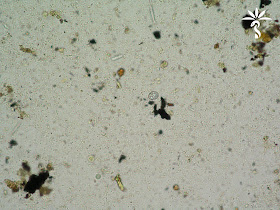
It's the first Monday of the month, and time for our case from Idzi Potters and the Institute of Tropical Medicine, Antwerp. The following objects were seen on a wet mount (with iodine) of a concentrated stool specimen (400x original magnification).Size is between 15 and 20 µm in length.
Here is another view on a carbolfuchsine staining according to Heine (1000x). Identification?
Here is another view on a carbolfuchsine staining according to Heine (1000x). Identification?
Sunday, February 3, 2019
Answer to Case 530
Answer to Case of the Week 530: Sarcocystis sp.; either S. hominis or S. suihominis
Thanks again to Idzi Potters and the Institute of Tropical Medicine, Antwerp, for sharing this case with us. I haven't featured intestinal sarcocystosis on this blog before, and so having these great photographs is a real treat. As Old One, Florida Fan, Blaine and others noted, humans serve as the definitive host for the intestinal form of sarcocystosis and shed sporulated oocysts and sarcocysts in their stool. Both forms are seen in this case:
Oocysts contain 2 sporocysts. Due to their fragile nature, they easily rupture so that free sporocysts are also commonly seen in stool specimens. Humans acquire S. hominis and S. suihominis by ingesting undercooked beef or pork respectively. As a pathology resident, I performed a survey of U.S. beef in collaboration with the USDA and confirmed the results of previous surveys that S. hominis was not present in the U.S. beef supply. Having said that, I still wouldn't recommend eating raw beef! Most cases of intestinal disease are asymptomatic, but infected individuals may experience mild watery diarrhea, fever and chills.
Rarely, humans may also serve as the intermediate host for some Sarcocystis species when ingesting oocysts or sporocysts in contaminated food or water. In this form of disease, sarcocysts form within various muscles in the body, causing transient myalgias, muscle weakness and associated edema. HERE is what a sarcocyst looks like in skeletal muscle (a case of S. cruzi; not a human pathogen).


