1. Embed arthropods and worms in casting resin, using products purchased at your local arts and crafts store. I've used Castin' Craft in the past, but there are other options out there as well. Here is one of my creations - adult Ixodes scapularis ticks (in different stages of engorgement) in a small petri dish:
Embedded arthropods are great for teaching because they are durable (e.g. their legs don't fall off from being manipulated over and over again), and you can easily examine both their dorsal and ventral aspects. If you're feeling really creative, then you can also make them into little works of art - or even jewelry (start a fun new trend). You can buy molds in every shape and size under the sun.
2. Find some anisakids in frozen (or fresh) cod. I have the best luck with Atlantic cod; in every bag of 10 fillets, I usually find at least 1 worm. I let the fillets thaw in the refrigerator and then use blunt dissection to find the coiled larvae. I put the fish (with anisakid) on a plate for display in my teaching lab. It's a great way to get medical students, residents, techs, and clinicians interested in parasitology.
For added fun, have a fish dissecting party! Here are some of my awesome pathology residents, who came up with this idea all on their own.
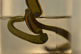
3. Buy some medicinal leeches (Hirudo medicinalis) at your hospital pharmacy. Like the cod, these also make a great display for a parasite teaching laboratory. You can use them as an example of an ectoparasite, talk about the anticoagulant they produce (hirudin - used medicinally), discuss their fascinating history (past and present) in medicine, and also discuss why it is necessary to give prophylactic antibiotics to recipients of leech therapy (to prevent sepsis with Aeromonas - a commensal bacterium in the gut of medicinal leeches).

Yes, it's a display with shameless 'ick' factor, but it's great for capturing the attention of medical students and clinicians alike.
4. Learn some basic entomology skills. Try dragging for ticks, collecting black fly (Simulium sp.) larvae from a fast-flowing stream (below), or learn how to mount insects professionally for teaching purposes (compliments of Blaine Mathison).
5. Last, but not least, have a parasite potluck, with treats decorated with your favorite (candy) parasites.
Cupcakes by Dr. Rachael Liesman, my Clinical Microbiology fellow:
Malaria cookies by my Education Specialist, Emily Fernholz
Cakes from the Mayo Medical Students! Coconut rectum, Giardia duodenalis, Reduviid bug transmitting Trypanosoma cruzi through its feces, Strongyloides stercoralis on a stool agar culture, and a strawberry cervix.


Coconut rectum is the appearance of a rectum laced with Trichuris trichuria worms in a heavy infection. The rectum may be prolapsed.
ReplyDeleteStrawberry cervix refers to a cervix with red petechiae from a Trichomonas vaginalis infection.
Your suggestions on mounting arthropods are very useful in a training setting. Most difficulties lie in the fact that the arthropods move around and do not co-operate with us,and if we euthanize them, they will only look dead. To overcome this, it may be useful to anesthetize them with CO2 prior to euthanization and mounting.
Florida Fan
Too fun and too clever - thanks x 400. Richard Garcia-Kennedy
ReplyDeleteYour arthopods look great, I never thought in using stuff from arts & crafts like resin and such, we use a mixture of Entellan with xylol to do something similar, but the result is not so smooth. We also use this mixture in wet mounts around the cover slip, they can be preserved for weeks if done right.
ReplyDeleteJust like Florida Fan said, arthopods don't cooperate to be seen by students, specially eyes from Loxosceles laeta, so we put them in the freezer for 15 minutes, it's the best way we found to authanize them and preserve its morphology prior to fixation.
-HLCM fan.
congrats on your 400th post Bobbi, I followed this blog with great joy and will do so in the future.
ReplyDeleteHans Naus from the Netherlands