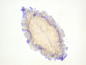
This week's case was generously donated by Dr. Julie Ribes. The following objects were seen in a Papanicolaou-stained urine specimen from an elderly man with hematuria. They varied in size, measuring ~ 70 micrometers in length. All images were taken using the 40x objective.
Identification?


I vote for some sort of crystal.
ReplyDeleteUric acid crystals.
ReplyDeleteI've got no experience with pap stain, but I agree: uric acid crystals.
ReplyDeleteThe pictures give me the appearance of crytals aggregates forming possibly around a uric acid nucleus.
ReplyDeleteFlorida Fan
I also agree with my predecessors. They look like uric acid crystals. Too small to be S. haematobium eggs, although they can sometimes lead to confusion Parasite eggs in urine cytology: Fact or artifact?
ReplyDeleteI also agree. The variation in size, along with the distinct lack of any anatomical features, points me in the direction of a crystal of some sort, likely uric acid due to the diamond shape. The edges of the structure appear "sharp", even ragged, particularly in the third photo. The delicate and "fragile" appearance of the object in the last photo is especially unique, and I would not expect to see anything of the sort in an ova, cyst, or parasite.
ReplyDeleteI was also thinking uric acid crystal
ReplyDeleteArtifact of some sort, likely crystal. I wouldn't try to identify crystal type on this particular preparation.
ReplyDeleteUric acid crystals from morphology and size
ReplyDeleteI doubt it a Schistosoma haematobium ova surrounded by many crystals.
ReplyDelete.....And the correct answer is?...
ReplyDeletelook like uric acid crystals
ReplyDelete