This week's case is generously donated by Idzi Potters and the Institute of Tropical Medicine, Antwerp.
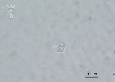
The following were seen in used contact lens solution from a young woman with complaints of eye pain and blurry vision. The first two images are taken with light microscopy, and the third with phase-contrast microscopy. What is your diagnosis? Please describe the forms you are seeing.


This a typical case of Acanthamoeba keratitis. Beautiful images from a Cyst and trophozoites.
ReplyDeleteAcanthamoeba cyst and troph
ReplyDeleteWow, pictures perfect rendition of the “thorny” amoeba and its polygonal cyst. Thank you Idzi for the classic case.
ReplyDeleteFirst picture - double walled Cyst, inner wall polygonal in shape - Acanthamoeba cyst
ReplyDeleteSecond and third - the thorny appearance of the Acanthamoeba trophozoite
-CJW-