This week's case just came through our lab this morning! These peripheral blood specimens are from a 30 year old male, originally from South Africa, who presented to the Emergency Department after several days of fever. He was emergently treated and is fortunately doing well now.
Diagnosis?
Tuesday, June 23, 2015
Sunday, June 21, 2015
Answer to Case 354
Answer: Plasmodium falciparum infection
Lots of great discussion on this case! This was the highest percent parasitemia I have seen in a non-fatal case of malaria. It was calculated at 24% in our lab. Fortunately this rapidly dropped with IV artesunate therapy and the patient is now doing well.
The key diagnostic features of this case were:
1. Heavy parasitemia, involving all stages of RBCs (note that some reticulocytes were infected). Plasmodium vivax and P. ovale only infect reticulocytes, whereas P. malariae is restricted to older RBCs; for these reasons, infections with these species will not reach the high level of parasitemia seen in this case.
2. The predominant form seen is delicate rings with 1-2 chromatin dots. Usually only rings (early trophozoites) and, less commonly, crescent-shaped gametocytes are seen with P. falciparum infection. However, this was a special case in which schizonts containing 8-24 merozoites were also seen. This is consistent with a very heavy infection in which the later stage forms that are usually sequestered in the peripheral capillaries have spilled out into the peripheral blood.
Thick blood film:
Thin blood film:
The main differential diagnosis, as pointed out by Lee, is babesiosis, which also consists primarily of delicate ring forms and can present with very high parasite loads. However, babesiosis can be excluded by the presence of schizonts and malaria pigment (hemozoin).
Finally, as a bonus, I saw a few of the following structures which I think might be microgametes. This is a stage usually only seen in the mosquito, but can rarely be seen when microgametocytes undergo a process called exflagellation and release microgametes (male gametes). See if you agree with me!
Tuesday, June 16, 2015
Case of the Week 353
This week's case was generously donated by Bryan Schmitt and Stephanie Kinney at Indiana University. Several specimens measuring approximately 1 cm in length were obtained during routine colonoscopy on a hispanic woman in her 50's; they were mistaken as colon polyps and submitted for histopathology. Identification?
H&E, 20x
H&E, 400x

H&E, 20x
H&E, 400x

Sunday, June 14, 2015
Answer to Case 353
Answer: Taenia saginata proglottid
There are several key morphologic features that allow for identification of this specimen. First, the presence of loose, 'myxoid' stroma, thick acellular cuticle and calcareous corpuscles allow for general identification as a cestode; in this case, a proglottid.
Next, identification of the small eggs with radial striations allow for further confirmation that this is a cestode, and identification to the Taenia genus.
Finally, the species can be tentatively identified as T. saginata, based on the presence of >13 primary branches (arrow heads) off of the central uterine stem (arrow).
Florida Fan noted that T. asiatica also has > 13 primary uterine branches and is essentially indistinguishable from T. saginata. However, Arthur rightly mentions that T. asiatica has not been described outside of Asia, presumably due to somewhat unique culinary habits of some Asian populations that include consumption of raw pig liver. Given that this patient was from Central America, she likely did not have exposure to T. asiatica, although we would need to perform molecular testing on the specimen to definitely rule out this species.
Sunday, June 7, 2015
Case of the Week 352
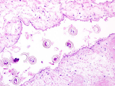
The following structures were seen in a hematoxylin and eosin-stained perianal skin biopsy from an Ethiopian man with recurrent colorectal fistulae and abscesses. Presumptive identification? What study would you recommend to confirm your diagnosis?
100x total magnification

400x total magnification

100x total magnification
200x total magnification

400x total magnification

Saturday, June 6, 2015
Answer to Case 352
Answer: Schistosoma sp. ova; most likely S. mansoni
As the readers pointed out, the appearance of eggs within granulomas in a perianal skin biopsy is highly suggestive of schistosomiasis, and image 4 might have the beginning of a lateral spine to support the diagnosis of S. mansoni infection. Unfortunately, the eggs tend to collapse in tissue sections and therefore it is difficult to definitively identify the characteristic spines. A stool examination is recommended to confirm the identification of these eggs. In this case, the O&P confirmed that this was S. mansoni infection.
Thank you all for writing in!
Monday, June 1, 2015
Case of the Week 351
Greetings from the New Orleans, the site of the 2015 ASM general meeting! This week's case is a conventional endocervical smear stained with Papanicolaou stain. The images are taken at 400x total magnification, with the orange-staining structures measuring approximately 60 micrometers in length.
Identification?
Identification?
Subscribe to:
Comments (Atom)


