This week's impressive case was generously donated by Dr. Ryan Relich. The following was seen in Giemsa-stained thick and thin blood films (1000x) from a middle aged immunocompromised male with recent travel to Niger. Diagnosis?
Monday, February 20, 2023
Sunday, February 19, 2023
Answer to Case 711
Answer to the Parasite Case of the Week 711: Plasmodium falciparum infection with high parasitemia. Primarily early stage trophozoites are seen, but a single (somewhat) banana-shaped gametocyte and possibly later stage trophozoites are often seen. Given the high level of parasitemia, it's not surprising that we are seeing some some extracellular forms. If there happens to be concern about an alternate diagnosis of babesiosis (i.e., if the travel history wasn't known), then PCR could be performed. In this case, the presence of hemozoin and elongated gametocytes allows us to rule out babesiosis from the differential diagnosis.
Florida Fan nicely describes this case as follows:
"The infected red cells are of all sizes; this shows that there’s no predilection either for young red cells or older red cells." (There are also no Schüffner's dots). "As such we can rule out P. vivax, ovale and malariae. The multiple infection with double rings, appliqué forms, and head phones orient us further into an identification of P. falciparum. The lone banana-shaped gametocyte further confirms P. falciparum being the causative agent. There is no morphology forms (or travel history) to indicate a possible monkey malaria. With such a high parasitemia this case is serious cause to emphasize the urgency that all malaria testing are to be a stat test 24/7."
Indeed, all cases of suspected malaria should be evaluated on a a STAT basis given the potential life-threatening nature of infection.
Here are some of the key identifying features of this case:
Monday, February 13, 2023
Case of the Week 710
This post is in recognition of Valentine's day while also keeping in our theme of parasites in muscle. The organ of interest this week is the heart of course! The following objects were seen in a endomyocardial biopsy from a patient with unexplained heart failure. What is your diagnosis?
Sunday, February 12, 2023
Answer to Case 710
Answer to the Parasite Case of the Week 710: Trypanosoma cruzi amastigotes forming a pseudocyst in cardiac muscle. Note that you can make out the nucleus and kinetoplast of some of the amastigotes:
While I posted this on Valentine's day, Chagas disease is actually a story of heartbreak. Here is a rather poignant poem about Trypanosoma cruzi by @DrCindyCooper:Fortunately treatment can be effective when the disease is caught early. April 14th is World Chagas Disease day, where the WHO and partners raise awareness pf this "silent disease" that affects mainly poor people with limited access to health care.
Monday, February 6, 2023
Case of the Week 709
Since we are on the theme of parasites in muscle, here is another case from my archive. What parasite is shown here?
Sunday, February 5, 2023
Answer to Case 709
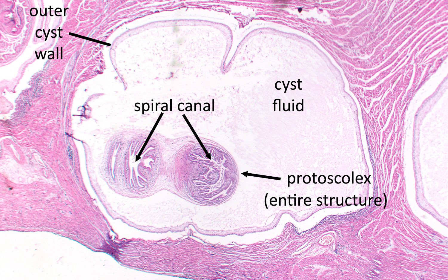
Answer to the Parasite Case of the Week 709: Taenia sp. cysticercus (larval form) in muscle. Since this case was from my archives, I don't know the source of the muscle (i.e., human vs. cow); however, given the number of cysticerci present and the co-existence of Sarcocystis sp. sarcocysts (not shown well, but you can see them if you enlarge the two images), I'm guessing it was a bovine case. Regardless of the origin, the features seen here are characteristic for cysticerci, including the overall shape and size, and the appearance of the internal protoscolex. Note the spiral canal which is often one of the clearest identifying features of the protoscolex.


