Here is our last case of 2024, courtesy of Drs. Courtney Comar and Laura Lamps. The following structures were seen in tissue removed during surgery from the peritoneal cavity of a patient with volvulus of the bowel and associated inflammatory changes. On histopathology, numerous foreign body giant cells and 'pulse granulomas' were seen. What are these structures?
Monday, December 30, 2024
Sunday, December 29, 2024
Answer to Case 764
Answer to the Parasite Case of the Week 764: Artifact; not a human parasite. This material closely resembles plant spirals. This was a challenging case to be sure! However, there were a few clues that can help us out here:
First, note that the outer contour of these worm-like objects consists of spiral, refractile rings that look a bit like a 'slinky' (for those of you who remember those old toys!)
What appears to be internal nuclear material is just multinucleated giant cells from the host.Second, I mentioned that numerous foreign body giant cells and 'pulse granulomas' were seen on histopathology. Pulse granulomas are granulomas associated with a foreign-body reaction and often contain identifiable vegetable material. The term 'pulse' refers to the edible seeds of leguminous vegetables. The presence of vegetable material elsewhere in this specimen support our diagnosis of vegetable spirals here.
For further confirmation, I turned to our friend and botany expert, Dr. Mary Parker, for her advice on this case. She indicated that we are likely seeing xylem vessels with spiral thickenings from plant vascular bundles (veins). She noted that "the simple spiral thickening in the above is typical of plant material such as young leaves (i.e. not as heavily thickened as in woody material), whereas the xylem thickenings in the colon wall are much more heavily lignified, indicating more mature plant material." She also explained that "young leaves like spinach, lettuce and parsley will all have a mixture of lightly and more-heavily thickened xylem." I remember learning about xylem and phloem back in primary school, but I never thought they would be useful in parasitology. Many thanks to Mary for her input, and kudos to Idzi for getting this case correct!
Check out the beautiful xylem spirals from this POST.
Wishing you all a very happy, healthy, peaceful, and productive new year.
Monday, December 16, 2024
Case of the Week 763
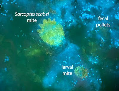
This week's case was donated by Dr. Andrew Bryan, Molly Weatherholt, Clarissa Ljungren, Bashi Ebrahimi, and Jacob Karsten. These striking images are from skin scrapings from an elderly patient with suspected superficial cutaneous fungal infection. The material was stained with Calcofluor white and examined with fluorescence microscopy. Diagnosis? What are the objects seen here - the big and little ones?
Sunday, December 15, 2024
Answer to Case 763
Answer to the Parasite Case of the Week 763: Sarcoptes scabei mites and their scybala (fecal pellets) - or, as Mike M. said - "scabies, and a lot of their poop!"
As Idzi noted, "...the small stuff on the 2nd image has the right size and the typical “puck”-shape with a dent, to be compatible with mite-droppings." However, Florida Fan also mentioned that we need to rule out small yeasts such as Malassezia spp. since they could also be present in the same slide - a great reminder!
In case you were wondering what scabies eggs looks like, here is a case that Florida Fan donated to the blog years ago:
Tuesday, December 3, 2024
Case of the Week 762
This week's case comes from my own lab - an unexpected finding on routine colonoscopy. These beautiful photos are courtesy of Felicity Norrie. Identification?
Sunday, December 1, 2024
Answer to Case 762
Answer to the Parasite Case of the Week 762: Enterobius vermicularis male
As noted by Idzi Potters, "The ribbed cephalic alae are typical for an Enterobius vermicularis adult. The curved tail and spicula (although retracted) point towards an adult male." These features can be easily appreciated in the below composite image. Thanks again to Felicity for the beautiful photographs!
Thanks also to all of those who wrote on Facebook, X (formerly Twitter), and LinkedIn with questions and comments. Some were intrigued that this worm was found on routine screening colonoscopy, which prompted me to count up the number of cases on my blog that had been discovered in this manner. It turns out that we multiple examples of adult nematodes and cestodes featured, including Trichuris trichiura (Cases 73, 155, 306, 362, 405, 448, 490, 515, 598, and 631), hookworm/Ancylostoma duodenale (Cases 520 and 759), Taenia saginata (Case 353), Rodentolepis (Hymenolepis) nana (Cases 756, 664, and 169), and Dipylidium caninum (Case 378). We also had a previous male E. vermicularis on this blog detected during screening colonoscopy (Case 576). I wasn't aware that I had accumulated so many of these incidental cases over the years!Others asked about the clinical significance of this finding. In this case, the patient was asymptomatic and there was likely no clinical relevance. This is not surprising, as enterobiasis is frequently asymptomatic,. However, it is likely that this patient was treated to eliminate the chance of him serving as a reservoir for infection.


