Monday, April 30, 2018

Answer to Case 492

Answer: Histoplasma capsulatum
This small oval-shaped fungus has a very similar appearance to the amastigotes of Leishmania species (and Trypanosoma cruzi), as well as the tachyzoites of Toxoplasma gondii. It is therefore always in my differential of small intracellular objects when I am looking at blood smears, bone marrow aspirates and tissue sections. All of these objects measure 2 to 5 micrometers in greatest dimension and can be found within phagocytic cells. However, there are several key differentiating features that allow for a correct identification to be made:

1. Leishmania spp. amastigotes - have a single nucleus and rod-shaped kinetoplast. They are found within macrophages/monocytes.
2. T. cruzi amastigotes - are indistinguishable from Leishmania amastigotes but have a different tissue tropism (e.g. cardiac and smooth muscle cells) and associated clinical presentation.
3. Toxoplasma gondii tachyzoites - are arc-shaped (but can appear oval in tissue) and have a single nucleus with no kinetoplast. They can infect any nucleated cell.
4. Histoplasma capsulatum yeasts - are found within macrophages/monocytes and divide by narrow-based budding. As Sugar Magnolia mentioned, their cell wall does not take up many stains well (e.g. Giemsa, H&E), and therefore they appear to have a surrounding capsule (sometimes termed a pseudocapsule). This is where the species name, capsulatum, comes from. A silver fungal stain (e.g. Gomori methenamine silver) will stain all of the yeast including its cell wall. In contrast, amastigotes and tachyzoites do NOT stain with GMS.

It's important to note that other yeasts such as Penicillium marneffei and Cryptococcus neoformans will also stain with silver fungal stains and may have a similar appearance to H. capsulatum in peripheral blood films. They can be differentiated through a careful examination of several morphologic features; Cryptococcus neoformans has a true capsule, unlike H. capsulatum, and exhibits more size variability, whereas Penicillium marneffei divides by formation of transverse septations rather than budding.

Whew! That was a long explanation. Kudos to my excellent parasitology technologists who correctly detected and identified the H. capsulatum on the malaria smears. I later found out that the patient was severely immunocompromised with AIDS - a common scenario for histoplasmosis detected on peripheral blood films.


Monday, April 23, 2018

Case of the Week 491

This week's case is courtesy of Dr. Delgado and the Yale Pathology department. The patient is young adult male with extensive travel throughout Europe, Central America and Africa, who presented fever, drenching night sweats, extreme fatigue, cervical lymphadenopathy and splenomegaly. CBC showed pancytopenia. The following are from the H&E-stained bone marrow biopsy and Giemsa-stained aspirate.


Identification?

Sunday, April 22, 2018

Answer to Case 491

Answer: Visceral leishmaniasis due to Leishmania species (Leishmania donovani complex or L. tropica). As noted by Brady Page, Anon, Florida Fan, William Sears, and Daniel Skipper, the diagnosis can be easily secured by identifying the amastigotes within mononuclear phagocytes. Each amastigote has a small eccentric nucleus and rod-shaped kinetoplast, giving the so-called "dot-dash" pattern (thank you @JClinMicro)
 And thank you to our poet, Blaine, who gave us the following:
Sheesh! This guy has Leish! 
Maybe from sandflies at the beach? 
PCR should be within reach, 
in case there is a call for Pentostam to release!

Monday, April 16, 2018

Case of the Week 490

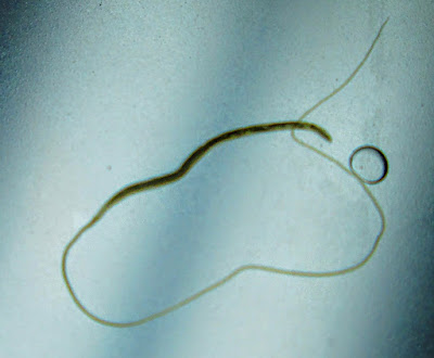
This week's case is generously donated by Florida Fan. The following object was submitted to the laboratory for identification. It had been retrieved during colonoscopy from the cecum of a 40-year-old man.


Identification?

Sunday, April 15, 2018

Answer to Case 490

Answer: Female Trichuris sp.
The most likely etiology is T. trichiura. However, as several readers mentioned, human infection with T. vulpis has rarely been reported and the adult worm has a similar appearance. The two worms are differentiated primarily by their eggs, with T. vulpis eggs being nearly twice as large as T. trichiura eggs. The only eggs we see in this case are immature and intrauterine; therefore it is not possible to use the eggs to help us differentiate the two worms.

Here are some of the key diagnostic features of this case:
Note that the head is at the slender end (all the better for embedding into the large bowel mucosa), while the larger end (containing the uterus and eggs) hangs free in the bowel lumen. We can tell that this is a female worm because of the eggs in the uterus (below) and because the tail is not coiled like a male's tail would be.


Monday, April 9, 2018

Case of the Week 489

Happy Unicorn Day! (yes, there really is such a thing).
This was seen in a trichrome-stained stool specimen. It measures approximately 19 micrometers in diameter, not including the 'horn'.
These were also seen in this specimen:


Identification? Images are courtesy of Emily Fernholz, MLS(ASCP).

Sunday, April 8, 2018

Answer to Case 489

Answer: Entamoeba coli or 'uniamoeba'!
While this case shows a beautiful trophozoite 'unicorn' for national unicorn day, it has an atypically-large karyosome which makes it challenging to identify it as E. coli. I therefore added some additional trophozoite photos from this case. Note the smaller, eccentric karyosome and clearly clumped peripheral chromatin seen in the following trophs:
As Idzi pointed out, this one looks like a dog's head!
The cysts were clearly consistent with E. coli, containing >4 nuclei (8 in the mature cyst). 

Monday, April 2, 2018

Case of the Week 488

Happy April! It's time for our first case of the month by Idzi Potters and the Institute of Tropical Medicine, Antwerp.

The patient is an adult male without known travel history who passed the following worm-like structure. It was placed in physiological saline solution and sent to the laboratory for identification.


In the lab, a drop of the saline solution is also examined microscopically, revealing the structure in the picture (size: 32 µm).


Diagnosis please?

Sunday, April 1, 2018

Answer to Case 488

Answer: Taenia sp.
Molecular testing showed this to be Taenia saginata.

There was a lot of great discussion regarding the differential on this case. Some of the key features were as follows:
1. Large size - although not given, you can see that this is a large tapeworm that fills the Petri dish. Even if this was a small Petri dish, the size would be most consistent with Taenia spp., Diphyllobothrium spp., and possibly Hymenolepis diminuta.

2. Size/shape of proglottids - this is one area where the ID gets tricky. Mature proglottids of H. diminuta and Diphyllobothrium are wider than they are long, whereas mature proglottids of Taenia spp. are longer than they are wide. The caveat is that immature proglottids of Taenia (as seen in this case) are also wider than they are long. Thus all 3 of these worms are in the differential. However, the proglottids of H. diminuta are very short and not like the ones shown in this case. Therefore, this diagnosis is less likely, leaving us with Diphyllobothrium and Taenia species as considerations.

3. Internal proglottid structures - this is a very helpful feature of this case since an internal 'rosette-shaped' uterus (consistent with Diphyllobothrium) is NOT present, but instead the opening to the lateral genital pore can be seen; the latter is consistent with this being a Taenia sp. proglottid.

Taenia sp. (this case); arrows show openings to lateral genital pores:
Diphyllobothrium sp. shown for comparison showing central rosette-structured uterus:
H. diminuta showing extremely short proglottids (from CDC DPDx):
4. Morphology of the eggs - another tricky factor here! This egg clearly contained a hooked-onchosphere, but it lacked the outer shell of Hymenolepis and the outer striated wall of Taenia spp. As mentioned above, this was a Taenia sp., and thus the lack of the thick striated outer shell can be attributed to the immature state of the egg (matching the immature proglottids).