Saturday, March 26, 2016

Case of the Week 390

The week's case was generously donated by Blaine Mathison. Below are H&E-stained sections of a perineal abscess of a patient diagnosed with Fournier's gangrene with bacterial infection. Identification?
40x
 100x
 400x
 1000x


Friday, March 25, 2016

Answer to Case 390

Answer: Blastocystis hominis


This was a very unusual and interesting case which generated a lot of great discussion. Thank you for the comments! Blaine (who donated this case) and I both believe that this case represents contamination/colonization of pre-existing fasciitis with B. hominis in a patient with Fournier's gangrene, rather than primary infection with this parasite. Fournier's gangrene is an extensive and rapidly-progressive polymicrobial fasciitis often seen in the setting of impaired host response (e.g. diabetes) and is usually precipitated by local trauma. The necrotizing process commonly originates from the anorectal tract, which might explain how B. hominis came to be involved.


This case gives us a rare opportunity to see the classic vacuolar forms of B. hominis in H&E-stained tissue sections. They have a very similar appearance to what we see in stool exams, with a large central vacuole and peripheral nuclei.


Monday, March 14, 2016

Case of the Week 389

This week's case was generously donated by Dr. Eric Rosenbaum who received the following in his laboratory. Identification?


Sunday, March 13, 2016

Answer to Case 389

Answer: Human Bed bug (Cimex sp.)

This case shows the characteristic features of a bed bug, including the flattened body, brown-red color, segmented abdomen, vestigial wings (wing pads), and setae (hair-like structures covering the body).


The 2 species of bed bugs that are most commonly implicated in human infestations are Cimex lectularis, the common bed bug, and C. hemipterus, the tropical bed bug. Rarely, humans can also be incidental hosts of Cimex species of bats and birds, especially when the natural hosts are in or living near human dwellings. It is important to differentiate human and zoonotic bed bugs, since the management is usually different. The best way to do this is by examining the length of the setae. Human bed bugs have setae that are shorter than the width of the eye, while bat and bird bed bugs have setae that are longer than the width of the eye. You can appreciate this somewhat in this case:


Sunday, March 6, 2016

Case of the Week 388

This week's case is from Emily F. in my lab. No history is available.


Identification?

Saturday, March 5, 2016

Answer to Case 388

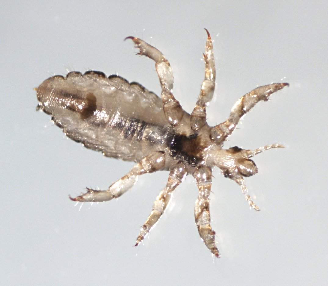
Answer:  Pediculus humanus, not further specified.

As several readers mentioned, knowing the source of the infestation would have been very useful in determining if this is a head or body louse, since these 2 subspecies (P. h. humanus and P. h. capitis) are morphologically indistinguishable, and overlap in size range (although head lice are on the smaller end of the range). Unfortunately, as is often the case, we were not given a source.

Pediculus humanus can be identified by its elongated body and grasping (raptorial) claws as shown below.


Sunday, February 28, 2016

Case of the Week 387

This week's case was generously donated by Dr. Heidi Lehrke. These objects were seen in a lymph node aspirate specimen.

May-Grunwald-Giemsa, 600x
May-Grunwald-Giemsa, 1000x


Identification?
How did they get into the specimen?

Saturday, February 27, 2016

Answer to Case 387

Answer: Giardia duodenalis (a.k.a. G. intestinalis, G. lamblia) trophozoites

This Giemsa-based stain nicely highlights all of the diagnostic features of Giardia trophozoites, including the flagella, which are not usually seen so well in trichrome or iron hematoxylin-stained stool preparations. The annotated image below shows some of the key diagnostic features:

Now to answer the important question that many of you asked - what are Giardia trophozoites doing in a lymph node?(!) Kudos to Anon, Florida Fan, and William Sears for correctly suggesting that the Giardia were a contaminant from the sampling method that was used.

What I didn't tell you is that the lymph node aspirate was performed using ultrasound-guided endoscopic biopsy. An endoscope was passed down the patient's esophagus, through their stomach and into the duodenum. Then, an aspirating needle was threaded through the endoscope and was used to take a sample of a neighboring lymph node by passing through the duodenal wall. Mystery solved!

This is the second case that I've seen of giardiasis detected incidentally during this procedure in the past 10 years, so it's important to understand how specimens are obtained.

Thank you for all of the great comments on this case. And thanks again to Dr. Heidi Lehrke for donating these beautiful pictures.

Saturday, February 20, 2016

Case of the Week 386

This week's case was discovered by the awesome techs in my lab while examining a concentrated wet-preparation from a stool specimen submitted for routine parasite exam. This is the only one of these objects identified. No further history was available.

100x magnification

400x magnification

Identification? What is the significance of this finding?


Friday, February 19, 2016

Answer to Case 386

Answer: Mite, not further identified.

It is difficult to identify this mite to the genus level based on the images shown. However, it is clear from the images that this is not Sarcoptes scabei or Demodex spp., two well-known human mites. Therefore, we are left with several options for identification:
1. This is a dust mite that was inadvertently swallowed.
2. This is a cheese mite or other mite commonly found in food.
3. This is a zoonotic mite that was accidentally ingested (none of which cause intestinal infection)

Therefore, this can be considered an incidental finding without clinical significance.