This week's case came through my lab awhile back and was beautifully captured in the following photographs by Emily Fernholz. The specimen is a Giemsa-stained thin blood film from a patient with travel to Botswana. What parasite is seen here?
.jpg)
.jpg)
.jpg)
.jpg)
.jpg)
.jpg)
.jpg)
This week's case came through my lab awhile back and was beautifully captured in the following photographs by Emily Fernholz. The specimen is a Giemsa-stained thin blood film from a patient with travel to Botswana. What parasite is seen here?
Answer to Parasite Case of the Week 677: Plasmodium malariae with classic band and schizont forms. There is also a hint of a basket form, and a nice collection of merozoites that recently burst out of an infected red blood cell. Note that the infected cells are smaller than the uninfected neighboring cells, which is classic for this species. Thank you for all of the great comments!


This week's case is generously donated by Dr. Adrienne Showler. The following was passed in stool by an 'adventurous eater', who regularly enjoys raw fish dishes, and also has tried raw beef and pork dishes in the United States. He reports passing similar objects on 4 separate occasions, and the length of each was between 1 and 3 feet. Unfortunately he didn't collect the samples (!), so we only have these photographs:


Likely identification?
Answer to the Parasite Case of the Week 676: Cestode, most likely Diphyllobothrium/Dibothriocephalus/Adenocephalus species. The CDC DPDx webpage on diphyllobothriasis explains how some of the names have changed for this group of tapeworms.
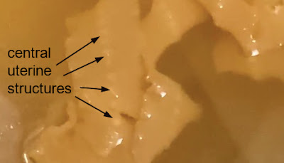
Unfortunately the specimen was not submitted to the laboratory and we therefore can't fully examine it. However, central uterine structure seen in the photo below is consistent with an adult tapeworm of the family Diphyllobothriidae:
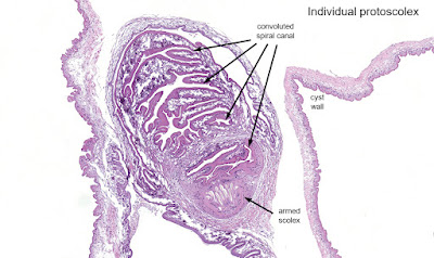
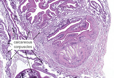
This week's case was donated by Dr. Paul Valenstein, and it is a beautiful example of an uncommon finding. It is an eyelid biopsy from a 6-year-old African child.

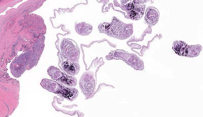


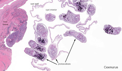
Answer to the Parasite Case of the Week 675: Coenurosis, a tapeworm infection with the larval form of coenurus-forming Taenia species, such as T. multiceps (the most commonly implicated species), T. serialis, T. brauni, and T. glomeratus.
Some of you noted the resemblance of this tissue infection with cysticercosis. That makes sense since human cysticercosis is due to the related cestode, Taenia solium. The primary difference is the presence of a single protoscolex in cysticercosis, vs. multiple protoscoleces in coenurosis.
Here is my approach to the diagnosis:
This week's case is courtesy of Dr. Charlie Huskins, one of our pediatric ID physicians. The parents of his patient found the following bug in their home and were concerned that it might be a disease vector. They are concerned that it might have bitten their child, as they have identified a potential 'bite' mark.
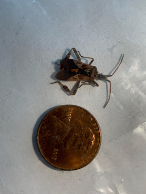
Of note, the family lives in the midwestern United States and has no history of recent travel.
What is the general identification of this bug? Is there any concern for disease transmission?
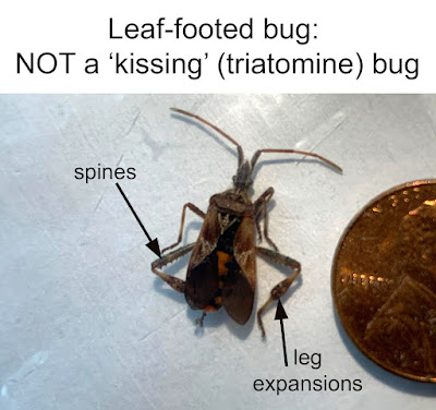
Answer to the Parasite Case of the Week 674: Not a human ectoparasite or disease vector; this is one of the Coreidae or "leaf-footed" bugs, most likely the western conifer seed bug (Leptoglossus occidentalis). The nickname comes from the leaf-like expansions on the legs of some species, usually on the hind tibiae. Leptolossus does not bite or sting, but can give off a pungent odor when disturbed. It feeds on sap and has long piercing mouthparts.
The importance of this case is that the specimen is a very convincing mimic of the triatomine vector of Trypanosoma cruzi - commonly called the "kissing bug." However, as several of you have pointed out, it can be differentiated by a number of key morphologic features such as the leg expansions and spines. The antennae are also much thicker than those of the triatomines. A hint to remember this from Khaled Itani - kissing bugs don't have spines on their legs. "If you want to be "kissing" you should be smooth." (I'll definitely be using this in the future!)
The comparative features are nicely described on the University of Wisconsin Department of Entomology page called "Is this a Kissing Bug" (thank you for the reference Luis, PJ Liesch, and Janet Waldron).
Based on this information, we can reassure our colleague and his patient that there is no cause for alarm with this bug. As PJ Liesch noted on Twitter, "It commonly sneaks into structures in the fall when seeking overwintering spots." Therefore, it is not surprising that our patient found this in her home in the midwestern US.
Welcome to Case of the Week 673, for this interesting day of two's (2/22/22 or 22/2/22, depending on your preferred format). To recognized this day, I've chosen a parasite to go with our theme of two's - histologic section of two parasites in lung tissue. You can click HERE to go to the digital image and view these parasites at higher magnification. Our non-pathologists may find the eggs seen on the digital slide to be quite helpful for making the diagnosis.

Answer to the Parasite Case of the Week 673: Paragonimus species; two adult flukes within lung tissue.
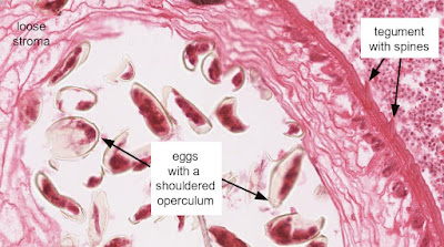
You can tell that these are flukes by their loose stroma, thin eosinophilic tegument (outer layer) with prominent spines, and suckers. As noted by Florida Fan, the characteristic eggs (each with a shouldered operculum), along with the location, are helpful in identifying this as a Paragonimus species. Excellent job!