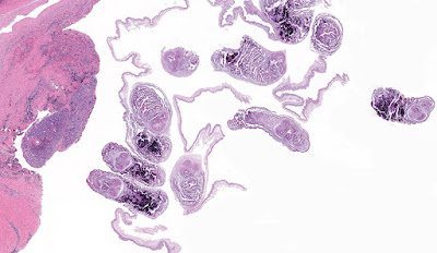
This week's case was donated by Dr. Paul Valenstein, and it is a beautiful example of an uncommon finding. It is an eyelid biopsy from a 6-year-old African child.
Diagnosis?
7 comments:
Anonymous
said...
The non staining hooklets of the invaginated scolex and the convoluted spiral canal indicated that this is a case of Cysticercus cellulosae. To tell the truth, I got it from the previous cases on the Blog. Thank you Dr. Pritt. Florida Fan
The multiple scoleces that seem to come from a unique germinative layer make me think to a case of coenurosis, due to Taenia serialis or Taenia multiceps.
At first sight I thought it's H nana but then I looked back and this case was from eyelids. Then I thought it could be cysticercosis. But some people on Twitter were saying case of myiasis. So much confusion....
Hi Unknown, Schisto can be ruled out by the fact that we can clearly see hooklets. Also the abundant presence of calcarous bodies indicates that a cestode is involved here! Interesting case for sure!
Every week I will post a new Case, along with the answer to the previous case. Please feel free to write in with your answers, comments, and questions. Also check out my image archive website at http://parasitewonders.com. Enjoy!
The Fine Print: Please note that all opinions expressed here are mine and not my employer. Information provided is for educational purposes only. It is not intended as and does not substitute for medical advice. I do not accept medical consults from patients.


7 comments:
The non staining hooklets of the invaginated scolex and the convoluted spiral canal indicated that this is a case of Cysticercus cellulosae. To tell the truth, I got it from the previous cases on the Blog. Thank you Dr. Pritt.
Florida Fan
The multiple scoleces that seem to come from a unique germinative layer make me think to a case of coenurosis, due to Taenia serialis or Taenia multiceps.
I agree with coenurosis - probably caused by T. multiceps as it involves the eye, but not excluding T. serialis as skin is involved as well (eyeLID).
At first sight I thought it's H nana but then I looked back and this case was from eyelids. Then I thought it could be cysticercosis. But some people on Twitter were saying case of myiasis. So much confusion....
Hmmm...I was thinking Schistosomiasis in the eye
Point well received Briceaut and Idzi.
Florida Fan
Hi Unknown,
Schisto can be ruled out by the fact that we can clearly see hooklets.
Also the abundant presence of calcarous bodies indicates that a cestode is involved here!
Interesting case for sure!
Post a Comment