Monday, August 29, 2016

Case of the Week 411

This week's case was generously donated by Florida Fan. The patient is a young African adult male. Shown are Giemsa-stained thick and thin blood films.


Identification?

Sunday, August 28, 2016

Answer to Case 411

Answer:  Malaria due to Plasmodium falciparum 

This case generated a lot of great discussion! As nicely described by Arthur V, there are "multiple infected RBCs containing 1 or more ring forms." Also, "the RBCs don't seem to be enlarged", "there are no amoeboid ring forms" and "the parasitaemia appears to be moderate judging by the number of ring forms present on the thick smear." These features, along with the absence of late-stage trophozoites and other intermediate forms are all consistent with P. falciparum infection. The following annotated images demonstrate some of the characteristic features seen in this case:

Thanks to everyone who wrote in on this interesting case.

Monday, August 22, 2016

Case of the Week 410

This week's case is in honor of the third annual Contact Lens Health Week (August 22-26). Contact lenses were received from a young adult male with bilateral conjunctivitis, photophobia and eye pain. These symptoms began shortly after he had been swimming in a freshwater lake while wearing his contact lenses. The contact lenses were plated for bacterial, mycobacterial, fungal and free-living amebic cultures, and fluid from the contact cases was sent to the cytology lab where it was made into a cell block. Here are photographs from the H&E-stained cell block specimen.


Identification?

Sunday, August 21, 2016

Answer to Case 410

Welcome to my new readers!

The answer to Case 410 is amebic keratitis due to Acanthamoeba sp.

As several of you mentioned, this organism can be identified by the presence of both trophozoites and cysts in the patient's specimen, as well as the characteristic source (ocular/contact lens) and exposure history (swimming in fresh water). Both Acanthamoeba spp. and Balamuthia mandrillaris form cysts and trophozoites in humans, whereas Naegleria fowleri only forms trophozoites (and rarely flagellated forms in cerebrospinal fluid), and therefore the latter can be excluded by the presence of the cyst forms. Of the two organisms that do form cysts in humans, only Acanthamoeba species are known to cause ocular infections - primarily in patients who wear contact lenses, thus giving us our final diagnosis.
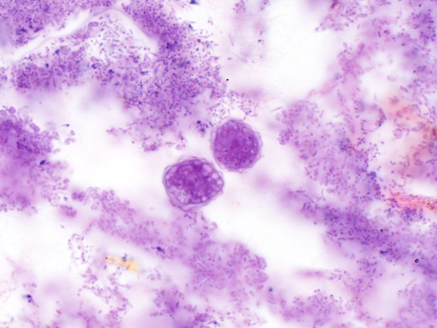
Trophozoite:


Cysts:
Acanthamoeba are free-living amebae that are ubiquitous in fresh water sources and can therefore easily contaminate contact lenses when worn during swimming. Note that there are also abundant bacteria present in this specimen. The bacteria that grew in the routine cultures from this patient were also common environmental organisms, and it is therefore they also likely were introduced while he was swimming with his contact lenses.

Monday, August 15, 2016

Case of the Week 409

Did you know that last week was National Public Radio's special series called Worm Week? You can check out their fun wormy posts HERE.

In keeping with the wormy theme, I thought I would revive one of my favorite cases from several years ago. I figured that the readers who only recently joined me might have not seen it, and it is definitely a good one. It was generously donated by Dr. Sandeep T.

The patient is a 27 year old woman with anemia and bilateral pedal edema extending to her knees bilaterally. She also reported a history of "vomiting a worm" so a colonoscopy was performed:

Some of the worms was removed for further examination:
Eggs were also seen in an ova and parasite exam from this patient:
Identification?

Sunday, August 14, 2016

Answer to Case 409

Answer: Fasciolopsis buski, the intestinal fluke

Although this is an older case (Case of the Week 227), I thought it was worth showing it again for the newer readers who may not have seen it. This is a parasite that we don't often see in the United States, but is common in Asia where freshwater plants are ingested.

The first step in making the diagnosis is recognizing that these objects are flukes - flat 'leaf-like' worms with an oral and ventral sucker:

The size of the fluke is also important. As you can see from the video, these flukes are quite large (!), measuring up to 7.5 cm in length. Intestinal obstruction and malabsorption commonly occur with heavy infections, as well as diarrhea, fever, abdominal pain and edema. The eggs are also important for making the diagnosis. In this case, thin-walled operculate eggs support our morphologic diagnosis.

Thank you to everyone who wrote in on this case!

Tuesday, August 9, 2016

Case of the Week 408

This week's case was generously donated by Dr. Carlos Chaccour. The patient is a young girl living in a rural region of  Venezuela who presented with a painful "pustule" on the back of her left thigh. The lesion had been present for the past four weeks.

Here is a video of the lesion:

Shortly after applying an occlusive ointment (in this case, soap paste), the parasite was easily removed from the wound:


Here is a closer view of the spines and mouthparts. 


Identification?

Monday, August 8, 2016

Answer to Case 408

Answer: Dermatobia hominis, the human botfly (3rd instar).

In this case, we see Dr. Chaccour applying an occlusive paste to 'suffocate' the botfly and facilitate its removal. Any type of thick paste will work - even bacon fat. Although we can't see the respiratory spicules, this fly larvae can be identified by the following features:

1. Robust (broad) body, often tapered at the terminal end (especially in earlier instars).
2. Concentric rows of spines on all but the terminal 3 body segments.
3. Found in Central and South America and the Caribbean
4. Causes furuncular myiasis (non-migratory boils)


Jon pointed out an excellent web page on this insect by the University of Florida. I'd encourage you to check it out - it has some beautiful images of the different instar stages.