Identification?
Monday, November 26, 2018
Case of the Week 520
This week's case is a small "worm" submitted for identification following removal during screening colonoscopy. It was tan-white and measured approximately 1.3 cm in length. The following are photos of its microscopic appearance:
Sunday, November 25, 2018
Answer to Case 520
Answer: Adult hookworm, Necator americanus
The hookworms are easily recognized by their piercing mouth parts - used for attaching to the small intestinal wall and drawing blood from the host. N. americanus has cutting plates, while Ancylostoma duodenale has sharp pointed teeth.
The hookworms are easily recognized by their piercing mouth parts - used for attaching to the small intestinal wall and drawing blood from the host. N. americanus has cutting plates, while Ancylostoma duodenale has sharp pointed teeth.
Thanks to all who wrote in with the great comments and ancient Chinese wisdom!
Monday, November 19, 2018
Case of the Week 519
Happy Thanksgiving day to my American friends! Here's a question for your consideration: which parasites can you get from eating undercooked turkey (the centerpiece of the classic Thanksgiving day feast)? Here is one of them in a squash preparation of a brain biopsy. What is it?
Giemsa, 100x objective with oil
Transmission electron microscopy:
Giemsa, 100x objective with oil
Transmission electron microscopy:
Sunday, November 18, 2018
Answer to Case 519
Answer: Tachyzoites of Toxoplasma gondii.
My accompanying request was to list other parasites that can be acquired from eating (undercooked) turkey. The two excellent responses I received from Bernardino Rocha were Gnathostoma spp. (likely, but we're not sure), and Trichinella pseudospiralis. Did we miss anything? Please write in if you can think of others.
Now, a few fun facts for the curious:
The tachyzoites are the rapidly-dividing form of T. gondii [tachy is from the ancient Greek ταχύς (takhús, “swift”)], and are the predominant form seen in acute and re-activated infections. In this case, the presence of numerous extracellular forms is evidence of an active infection. In contrast, tissue cysts containing bradyzoites, the slowly-replicating forms [brady is from the ancient Greek βραδύς (bradús, “slow”)] are seen during latent infection. The word zoite derives from the ancient Greek ζῷον (zôion, "animal"). I always make sure to point out word origins to my students when they are useful for remembering parasite names. For example, most medical students know the difference between tachycardia and bradycardia, and know to think of a 'zoo' as a place where animals are found. Helping them apply the knowledge they already have helps them learn these new, and often very foreign-sounding words.
T. gondii can infect any nucleated cell and tachyzoites are commonly seen within host cells during active infection. In this case, we can see both free and intracellular tachyzoites:
For those of you who like etymology, you may also be interested to know that the word Toxoplasma comes from the ancient Greek words τόξον (tóxon, "bow" or "arc") and πλάσμα (plásma, “something molded”); thus the name nicely describes arc-shaped form of this parasite. I love when parasite names actually make sense! You can especially appreciate this shape in air-dried touch preparations. Disappointingly, the arc shape is only rarely seen in sections of formalin-fixed, paraffin-embedded tissue, since the parasites tend to shrink and round up during fixation, taking on a more ovoid appearance. This makes it much trickier to differentiate them from small yeasts such as Histoplasma capsulatum and the amastigotes of Trypanosoma cruzi and Leishmania spp.
Transmission electron microscopy allows us to take a closer look at the T. gondii tachyzoites. As mentioned by Bernardino, the apical complex containing conoids and rhoptries is nicely seen:
HERE is an open access book chapter by the Global Water Pathogen Project that contains a reprint of the classic Dubey et al. 1998 figure showing the major structures of T. gondii tachyzoites and bradyzoites.
Monday, November 12, 2018
Case of the Week 518
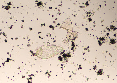
This week's case is a bit of a puzzle for you to put together. The following object was seen in a urine sediment. It was initially moving, but very quickly died. It measures approximately 130 micrometers in length.
Wet prep, 10x objective
Wet prep, 40x objective
Identification? Images are by one of our Clinical Microbiology fellows, Dr. Sarah Jung.
Wet prep, 40x objective
Identification? Images are by one of our Clinical Microbiology fellows, Dr. Sarah Jung.
Sunday, November 11, 2018
Answer to Case 518
Answer: Schistosoma haematobium miracidium, newly hatched with egg remnant.
This really neat photo, captured by our clinical microbiology fellow, Dr. Sarah Jung, nicely captured the miracidium right after it had exited the egg - AND - the egg remnant is still recognizable by its characteristic terminal spine. Great job Sarah!
This really neat photo, captured by our clinical microbiology fellow, Dr. Sarah Jung, nicely captured the miracidium right after it had exited the egg - AND - the egg remnant is still recognizable by its characteristic terminal spine. Great job Sarah!
I've tried to the best of my ability to label all of the components of the miracidium:
We rarely get to see these in the lab - although there is a hatching test you can try if you'd interested - and so it was a real treat to see the newly-hatched form of this parasite. Sarah mentioned that this was the best type of call to get called in from home for.
If you haven't already, I encourage you to go back and read the comments on this case - they are very interesting. I am so fortunate to have such a knowledgeable group of contributors who are always willing to share information and answer each other's questions. The comments also included a poem from Blaine which I will share here:
Humpty Haematobium sat on a wall
Humpty Haematobium had good fall
All the King's horses
and all the King's men
Couldn't put Humpty Haematobium back together again!
Excellent as always Blaine! Sarah and my lab staff have just a slight modification to your poem:
Humpty Haematobium sat in some pee
Humpty Haematobium yearned to be free
All of my fellows
Both women and men
Couldn't put Humpty together again!
Monday, November 5, 2018
Case of the Week 517
Case of the Week 513
This week features our monthly case from Idzi Potters and the Institute of Tropical Medicine, Antwerp.
A 45-year-old female patient, suspected of having an infection with Strongyloides stercoralis, provided a stool specimen for Baermann concentration. The following structure was found, measuring about 300 µm in extended state. Diagnosis please.
Sunday, November 4, 2018
Answer to Case 517
Answer: rotifer
Wow, great comments on this case! The Old One mentioned that this is a bdelloid rotifer. He comments "In this year of the women, it should be noted that bdelloid rotifers are all female. Able to be successful for millennia while maintaining genetic diversity by taking DNA from other creatures." Fascinating! According to the Encyclopaedia Britannica, "Rotifer, also called wheel animalcule, any of the approximately 2,000 species of microscopic, aquatic invertebrates that constitute the phylum Rotifera. Rotifers are so named because the circular arrangement of moving cilia (tiny hairlike structures) at the front end resembles a rotating wheel."
There is no clinical significance to this finding. Rotifers are found in environmental water sources, so it is likely that the organism entered the specimen through the collection process - possibly from toilet water contaminated with untreated water.
We've seen a rotifer before on this blog - in Case of the Week 304. Check out the photos from the case contributor, Ahrong Kim in South Korea - they're beautiful!
Friday, November 2, 2018
Halloween Parasite #5
HERE is Halloween Parasite #5, the last in my series of creepy dreadful wonderful parasites for the Halloween season. These pieces are written for the general public, including children, and aim to interest more people in the fascinating world of parasitology.
Thursday, November 1, 2018
Halloween Parasite #4
Happy Halloween Week everyone! As you all know, parasites can be creepy dreadful, but also fascinating, and sometimes even helpful. As a special Halloween treat, I'll be highlighting 5 different parasites on the Mayo Clinic News Network - 1 each day. HERE is parasite #4 - "worms in love" These are written as educational pieces for the general public. Feel free to use the text and images for your own educational purposes.
Subscribe to:
Posts (Atom)


