Sunday, December 31, 2017

Answer to Case 475

Answer: Chilomastix mesnili trophozoites.

Congratulations to Martin, Ali, Florida Fan, Mark, Atiya, Sara, and Alexandra who got this correct!

The videos show the beautiful 'spiraling' motility of this organism, similar but distinct from the 'falling leaf' motility of Giardia and the 'jerky' motility of Pentatrichomonas hominis. In the lab, of course, we would also have the final fixed morphology to aid in our diagnosis and confirm our impression from the direct preparation.

For comparison, you can view my (now very old) case of P. hominis at:
http://parasitewonders.blogspot.com/2008/01/parasite-case-of-week-5.html

And here are some beautiful videos of the falling leaf motility of Giardia by Idzi:


Monday, December 25, 2017

Case of the Week 474

Happy Holidays to all of my readers! This week's case is in the form of a Christmas tree. Can anyone tell what the tree is made out of? (hint: this is from a Trichrome-stained stool specimen)

Sunday, December 24, 2017

Answer to Case 474

Answer: Charcot-Leyden crystals, white blood cells and red blood cells

As Florida Fan mentioned, the C-L crystals are a guise for snow crystals, and the red cells may perhaps represent red delicious apples before man-made ornaments were innovated. I also envision the C-L crystals representing pine needles, perhaps?


I was very pleased to stumble upon this 'tree' when looking for some crystals to photograph. Other than the star, nothing else has been digitally added to this image.

Happy Holidays to all!


Monday, December 18, 2017

Case of the Week 473

The following structures were seen in a trichome-stained stool specimen. They measure approximately 10 to 20 micrometers in diameter. Identification?


Sunday, December 17, 2017

Answer to Case 473

Answer: Blastocystis sp. AND pollen.
Wow, you all impressed me by noting the less obvious Blastocystis in image 1, in addition to the pollen in all 3 images. Also, no one mistakenly thought that the pollen was a helminth egg (e.g. Taenia sp.) - a common pitfall. Excellent job!

While the Blastocystis cyst-like forms are the only parasites presents (image 1), I do agree that the pollen is the most striking finding. And very festive as Florida Fan and Idzi mentioned! I will defer the identification of the pollen to the others as this is outside of my area of expertise, but appreciate the time that some readers took to identifying the specific variety shown here.

Stay tuned for a holiday case next week.

Monday, December 11, 2017

Case of the Week 472

This week's case features an intriguing video by Dr. Graham Hickling.

The accompanying questions are:
1. What arthropod is shown here
2. What stages of the arthropod are seen?

Sunday, December 10, 2017

Answer to Case 472

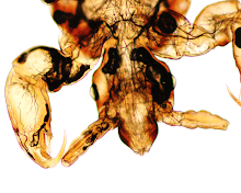
Answer: Hard tick (Ixodidae) nymph emerging from the larval exuvia. The inverted "U-shaped" anal groove on the ventral surface (photo below) allows us to identify this as an Ixodes species.
The gender is not possible to discern in nymphs, but as mentioned by Anon, it can be determined in adults by examining the scutum and basis capituli. The scutum (dorsal shield) of the female only covers a portion of the dorsal surface, compared to the male in which it covers nearly the entire dorsal surface. In the nymphal stage, both males and females have a similar-appearing scutum.  Also, only females have porose areas whereas males do not. The porose areas are located on either side of the basis capituli and produce antioxidants which are combined with waxy secretions from the Gene's organ. This substance is applied to eggs right after they are laid and serves as a protective coating. The porose areas also lubricate the Gene's organ, allowing it to expand and retract more easily.

Thanks again to Dr. Graham Hickling for donating this fascinating video. Thank you also to Dr. Robyn Nadolny for the additional information about the function of porose areas and Gene's organ. If you'd like to see the Gene's organ in action (and tick eggs being laid), check out this other amazing video by Graham.

Monday, December 4, 2017

Case of the Week 471

Welcome to the first case of the month featuring a case from Idzi Potters at the Institute of Tropical Medicine Antwerp. This case is really spectacular and something we don't see very often.

The following parasites were discovered in a man's peritoneal tissue during an inguinoscrotal hernia repair.  The resided in Benin, Africa. Here is the resected section of peritoneum along with the attached parasites (CLICK ON IMAGE TO ENLARGE):
 
Diagnosis?

Sunday, December 3, 2017

Answer to Case 471

Answer: Pentostomiasis; most consistent with Armillifer species. William Sears also suggested that Raillietiella sp. is also in the differential, which Idzi confirms. Both are found in African countries and associated with consumption of raw snake meat. Identification is accomplished morphologically by counting the annuli. You can also use molecular studies, although these are not widely available.

Idzi also provided me with the following beautiful (and very creepy) photos from his Institute's specimen archives which show an adult and larval Armillifer armillatus:

Idzi and his group published this case, so you can read more about it HERE. Fascinating case!