Monday, August 31, 2020

Case of the Week 604

 This week's case was generously donated by Dr. Peter Gilligan, and features a histopathology section of an appendix from a patient with Crohn's disease. The pathologist was concerned when they saw the following object in the lumen of the appendix. Identification?

Sunday, August 30, 2020

Answer to Case 604

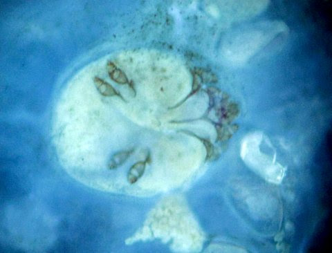
 Answer to Parasite Case of the Week 604: Plant material, not a parasite. Although the large size and round shape in section superficially resembles a cross-section of a large roundworm (i.e., Ascaris lumbricoides), there are some key features that allow us to identify this as plant material. 

As nicely stated by Florida Fan, "There does not seem to be a layer of polymyarian muscle cells as well as the other anatomical components such as a digestive tube or reproductive organs and no excretory organs either" which would be seen in Ascaris lumbricoides. He also adds: "when magnified enough we can see polygonal cells often observed on vegetable matters. The other observation is that there is a fine brown line inside which I believe to contain melanin pigment, a character we do not find in helminths."

These are excellent observations on the key features for distinguishing plant material - in this, likely a cereal bran - from round worms. The polygonal cells are a characteristic feature of plants, and can be seen in the outer layer of the structure in this case

This outer layer would likely have been birefringent with polarized light, which is another helpful feature for differentiating plant material from helminth body layers. Dr. Mary Parker, a botanist, kindly provided the following explanation for this case:  "I am pretty sure this is plant material, probably a few layers of the bran of a cereal grain. One of the layers of cereal bran contains a water-insoluble orange/red pigment called phlobaphene which give the grain its colour. This pigment layer is most obvious at 5, 8, and 12 o'clock on your section image. Botanists call a cereal grain a caryopsis because it is technically a seed with the fruit wall adhering to the outside (I explain this to my students as one pea in a pod with the pod adhering to the pea surface). Cereal bran therefore consists of layers derived from the fruit wall and the seed testa, and it is one of the testa layers that is pigmented. The digestive process does separate these layers." 

For comparison, here is a cross-section of A. lumbricoides, showing the thick acellular outer cuticle, underlying hypodermis, and tall polymyrian muscle cells. Note also the sections of internal organs.


Thanks again to Dr. Gilligan for donating this interesting case!

Tuesday, August 18, 2020

Case of the Week 603

 Here's a fun case for us this week from my technical specialist, Emily Fernholz. The following objects were seen on a Giemsa-stained thick film from a patient with recent travel to the Western United States. Diagnosis? What additional testing would you recommend?


Sunday, August 16, 2020

Answer to Case 603

Answer to Case of the Week 603: Borrelia sp. spirochetes. The history is most consistent with a relapsing fever Borrelia sp., and subsequent PCR showed this to be Borrelia hermsii

As Florida Fan mentioned, this case is a great reminder that when we examine a Giemsa-stained blood film, we may find things other than parasites. In addition to spirochetal bacteria such as in this case, we can find intracellular clusters (morulae) of Anaplasma phagocytophilum and Ehrlichica spp. bacteria, and intracellular years (e.g. Histoplasma capsulatum, Talaromyces marneffei). Leishmania spp. amastigotes can also rarely be seen within phagocytic cells. I've shown Borrelia spirochetes on this blog before (see Case of the Week 502), but never on a thick film, so I thought this would be a nice case to include here.

Note that the spirochetes are uniformly slender with broad undulations. This is different than the microgametes of an exflaggelated Plasmodium sp. microgametocyte which I previously showed on Parasite Case of the Week 595. In that case, I created a composite image of 'squiggly things' seen on a blood smear which may be helpful in identifying and differentiating them:

Some readers asked if the Borrelia species that cause Lyme disease could also be seen on blood film. The answer to this is NO - with one small caveat. The main cause of Lyme in the United States and Canada is Borrelia burgdorferi, whereas in Europe, you can also see B. garinii and B. afzelii. These organisms have extremely low levels of spirochetemia, and therefore spirochetes are NOT seen on peripheral blood films. The only rare exception to this is Borrelia mayonii, an uncommon cause of Lyme disease restricted to the upper Midwestern United States. This organism is actually found in much higher concentrations in the blood, and occasionally a rare spirochete can be seen on peripheral blood smears. We published on this several years ago; you can read the article HERE. (Let me know if you would like a copy).

There were many other good comments posted on this blog case - both in the Comments section and on Twitter - so I would encourage you to read them to learn more about the experiences from our readers.


Monday, August 10, 2020

Case of the Week 602

 This week's case was generously donated by Dr. Harsha Sheorey. The following object was submitted from a middle-aged man with hematuria. The specimen source is urine, and accompanying instructions asked the laboratory to rule out a fluke or cestode. The patient had traveled to Southeast Asia in the past.


Identification? What are the clinical implications for this patient?

Sunday, August 9, 2020

Answer to Case 602

 Answer to Parasite Case of the Week 602: insect larva, consistent with a member of the Psychodidae (drain/sewer fly) larva. As Richard Pollack noted, this is "Not from patient but from commode. Upon their discovery, folks often jump to the conclusion that they derived from the bladder or intestine. There are reports in the literature of myiasis from these flies, but one should generally be skeptical." It turns out that this is exactly what happened in this case. The specimen consisted of urine taken from the toilet water.

Florida Fan added "Being not an entomologist, I would say latrine fly larva and leave the entomology part to those specialists. The specimen has all the anatomy parts of water insect larva including a breathing tube at the posterior. For sure this would be a startling finding but, to the layman it may create a stunning effect at least."

I agree with Florida Fan that putting an actual name on this larva is not important for clinical care. What is important is identifying this as a non-parasitic larva. The presence of a respiratory siphon (breathing tube) in this case is a good indication that this is not a myiasis-causing fly larva. Other features seen here that would not be seen in a myiasis-causing larvae are bristles and a sclerotized head capsule.


Monday, August 3, 2020

Case of the Week 601

Here is our monthly case by Idzi Potters and the Institute of Tropical Medicine, Antwerp. The following were seen in skin scrapings from a young immigrant male. Identification?


Sunday, August 2, 2020

Answer to Case 601

 Answer: Sarcoptes scabei, the 'human itch mite'

As noted by Kosta, Patrik, and Anon, this is a larval mite, as evidenced by the presence of only 6 legs. Mites are arachnids and have 8 legs in their nymphal and adult stages. 

As noted by Old One in a previous post, Sarcoptes are round to ovoid when viewed from the back; when viewed from the side they are ventrally flattened and dorsally rounded (similar to a turtle). They possess short stumpy legs, and have no internal or external respiration apparatus (stigmata or tracheae). The ventral surface contains a number of chitinized plates called apodemes, the dorsal surface is partially covered by wide-angled, V-shaped-spines (>). The cuticular surface is sculptured into numerous parallel ridges which superficially resemble human finger prints, and the anus is at the posterior end of the mite (this is the characteristic used to differentiate Sarcoptes from Notoedres which has a dorsal anus and sometimes infests humans) It's dorsally-located anus makes it appear subterminal. Check out his additional comments in the comments on this case regarding Notoedres cati. This cat parasite can cause transient pruritic lesions in humans that closely resemble scabies.


Florida Fan also notes that S. scabei fluoresce in the traditional mycology calcofluor white preparation. Here are a few photographs he donated to the blog ages ago - simply stunning! 


Also, check out his beautiful photographs from Parasite Case of the Week 503: Demodex folliculorum, another mite found in human skin scrapings