Monday, June 27, 2022

Case of the Week 687

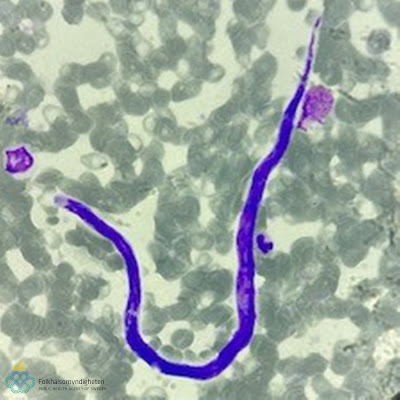
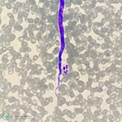
 This week's case is generously donated by Dr. Ioana Bujila of the Public Health Agency of Sweden. The patient is a 67 year old woman from Gabon. Blood was examined by direct mount and Giemsa-stained blood films, and the following were identified:


These objects are approximately 228 micrometers in length.

What is your diagnosis? Are there any additional laboratory analyses that are recommended in this case? 

Sunday, June 26, 2022

Answer to Case 687

 Answer to Parasite Case of the Week 687Loa loa

As noted by Florida Fan, @JuanCGabaldon, Idzi P, Ulrike E. Zelck, Priyanka Gupta, and others, the video clearly shows this to be a sheathed microfilaria, and the Giemsa smear shows the column of nuclei extending all the way to the tip of the tail, thus allowing us to make an identification of Loa loa. The patient's travel history (Gabon) also fits with this identification. 

As Idzi P. mentioned, I like to teach my students that nuclei flow-a flow-a to the tip in Loa loa - a fun learning aid!  Also check out this beautiful infographic by @cullen_lilley for an algorithm to differentiate the  common human-infecting microfilariae. 

Idzi P. further adds "These are by far the most beautiful images I have ever seen from what is definitely Loa loa microfilariae. I especially LOVE the video (second one) where the sheath is nicely visible!"

I agree - the video is amazing, and you should all check it out. 

Idzi P. goes on note that "a Knott's concentration could be envisaged to obtain a quantitative result before treatment is started. Also a hematoxylin-based staining (like Carrazzi's hematoxylin staining) could be used to demonstrate the sheath more nicely (as Luis H. points out: Giemsa does not usually/reliably stain the sheath of microfilariae) and better show the positioning of the nuclei in the tail." This is the answer to my question regarding additional laboratory testing that is indicated. Treatment of Loa loa is quite complicated, and patients with more than 8,000 microfilariae/mL are at risk of fatal encephalopathy with treatment. Therefore, obtaining a parasite count is important for guiding therapy. 

Thanks again to Dr. Ioana Bujila and the Public Health Agency of Sweden for sharing this beautiful case!
Tuesday, June 14, 2022

Case of the Week 686

This week's case was generously donated by Dr. Mike Mitchell and Laura Derderian. The following structure was seen in a stool concentrated wet mount specimen. It measures 27.5 micrometers in length. This was the only structure present in this specimen. Identification? What additional testing might also be useful in this case?


Sunday, June 12, 2022

Answer to Case 686

 Answer to the Parasite Case of the Week 686: Cystoisospora belli sporulated oocyst

This case shows a single - but classic - sporulated oocyst of Cystoisospora (formerly Isospora) belli. Note that two sporoblasts are present in this case, which is unusual to see in freshly passed stool specimens. Most C. belli oocysts are shed in an immature state - either unsporulated or partially sporulated (with only one sporoblast). They mature further in the environment, with the sporoblast dividing in two so that the mature oocyst has 2 sporoblasts. The sporoblasts become sporocysts with a surrounding cyst wall, and the sporocysts divide twice so that they produce 4 sporozoites each. 


We can make the diagnosis based on its characteristic appearance. However, if we wanted to confirm the diagnosis, Florida Fan notes that the oocysts "autofluoresce brightly under UV light excitation and will stain acid fast too." He goes on to say that "our team members prefer the autofluorescent methodology as it is simple and less time consuming." I know of some other labs that also prefer fluorescent microscopy to modified acid fast (MAF) staining since it is more sensitive. 

You can see some great examples of MAF staining HERE, and an autofluorescent oocyst of C. belli  from Florida Fan HERE.


Thanks again to Dr. Mike Mitchell and Laura Derderian for sharing this great case!


Monday, June 6, 2022

Case of the Week 685

 This is week's case is from our long-time contributor, Idzi Potters from the Institute of Tropical Medicine, Antwerp. The following structures were seen in a iodine wet preparation of concentrated stool. Identification? 


Sunday, June 5, 2022

Answer to Case 685

 Answer to the Parasite Case of the Week 685: Raphides, i.e, needle-shaped crystals of calcium oxalate or calcium carbonate used by several plants such as pineapple, kiwi, and rhubarb as a defense against plant-eating animals. According to Idzi, "The weird feeling you get in your mouth when eating too much pineapple is apparently caused by these crystals. You can find them quite often in stools and they should not be confused with Charcot-Leyden (C-L) crystals!" They can be differentiated from C-L crystals by their long needle-like shape and uniform diameter:

As a comparator, Idzi provided 2 nice photographs of C-L crystals, the slender bipyramidal hexagonal crystals of galectin-10. "They were first noticed in 1851 by Friedrich Albert von Zenker, and later on described in detail in 1853 by Jean-Martin Charcot and Charles-Philippe Robin. After that, in 1872, they were further described by Ernst Viktor von Leyden. Hence their name…"

Charcot-Leyden crystals:


Galectin-10 is contained in the granules from eosinophils (and also from basophils) and spontaneously crystallizes into Charcot-Leyden crystals when it is released in the extracellular environment (by exocytosis and/or cellular breakdown). Charcot-Leyden crystals are therefore indicative of eosinophilic inflammation (caused by asthma, allergic reactions, parasitic infections, etc)."

Thanks again to Idzi for this excellent case!