Sunday, July 31, 2022

Answer to Case 690

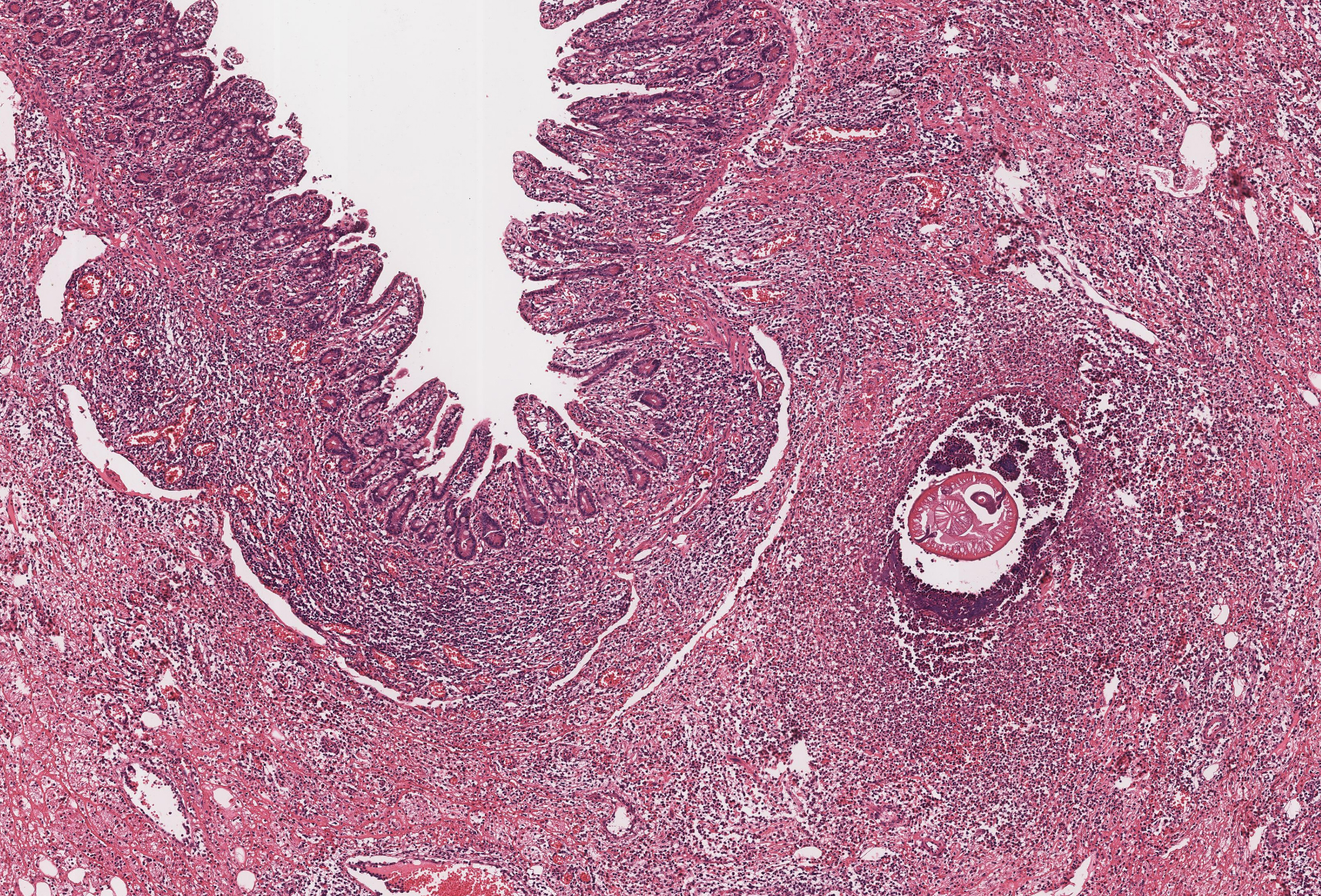
 Answer: Embedded hard tick, partially engorged with blood. 

As noted by Florida Fan, it is unfortunate that this tick was sectioned for histopathology, since "we need to examine the anal groove, the shield, the palpi, and the basis capitulum" in order to identify the tick to the genus and species level. This level of identification is helpful for patient management, since different species serve as vectors for different microorganisms. Additionally, finding an engorged Ixodes scapularis in a region of high Lyme disease endemicity may prompt antibiotic prophylaxis in certain situations. 

Without being able to examine the external features , we can only make some general comments about the tick in this case. As noted by Richard Bradbury, the tick is engorged, it has a scutum (dorsal shield), and the scutum takes up <1/3 of the body. Thus, we can state that this is a female hard tick. There is also an apparent lack of festoons (although this is hard to assess in sagittal section), and thus the tick may be an Ixodes species. Here are some of the key diagnostic features:

One may ask "why remove the tick like this at all?" Would it have been easier (and much less invasive) to simply remove the tick with tweezers? While I don't know why the tick was removed by excision in this case, I'm guessing that it was misidentified as a thrombosed skin tag. 

Thank you all for writing in with the excellent comments!


Monday, July 18, 2022

Case of the Week 689

 This week's case is from a middle-aged woman with unexplained small bowel perforation. Here is the section with the culprit. You can view the whole slide image HERE. What is your diagnosis?


Sunday, July 17, 2022

Answer to Case 689

 Answer to the Parasite Case of the Week 689:  Anisakid, most consistent with an Anisakis species.

Hopefully you all had a change to look at the whole slide image HERE. From its location in the small intestine, deep in the submucosa, one can envision how it may have extended through the wall of the intestine and caused a perforation. 

Dr. Luca Fanasca noted that "we can clearly see features consistent with an anisakid infection: multilayered cuticle (from a portion which seems detached, in the upper part), polymyarian muscle cells, the beautiful Y shaped lateral chords and the banana-shaped excretory cell. From the quite slender appearance of the lateral cords I think we could suppose this is actually an Anisakis sp., but a definitive identification would require the observation of other characteristics like the caecum position and shape, or molecular diagnostics strategies." 

Below are some of the key features of nematodes, and anisakids in particular: 

Features of all nematodes:

  1. Acellular outer cuticle (may be adorned projections in some nematodes (e.g., the lateral alae of Enterobius vermicularis, and the cuticular bosses of Loa loa)
  2. Underlying musculature. The musculature is either flattened (platymyarian) or tall (coelomyarian). The musculature is further categorized by the number of muscle cells: meromyairan (2 to 5 rows) or polymyarian (large number of rows in each sector)
  3. Lateral chords 
  4. Fully-developed intestinal tract
  5. Separate sexes (males and females)
Features of anisakids in particular:
  1. L3 larva embedded in the wall of the stomach or intestine (or in the peritoneal cavity)
  2. Thick unadorned cuticule
  3. Tall coelomyarian musculature (m)
  4. Splayed, Y-shaped lateral cords (arrow heads). The slender appearance of lateral cords are consistent with Anisakis species.
  5. Prominent esophagus (black asterisk)
  6. Banana-shaped excretory cell (white asterisk)


Tuesday, July 12, 2022

Bedtime Reading?

For those of you looking for some good (?) bedtime reading, you may be interested in two fun articles that Blaine Mathison and I recently wrote:

Blaine A. Mathison, Bobbi S. Pritt, Sleeping with the Enemy: Everything You Need to Know about the Biology, Clinical Significance, and Laboratory Identification of Bed Bugs. Clinical Microbiology Newsletter, Volume 43, Issue 1, 2021, Pages 1-7, https://www.sciencedirect.com/science/article/pii/S019643992030091X

Blaine A. Mathison, Bobbi S. Pritt, Don't Be a Nit Wit; Know Your Lousy Companions! Clinical Microbiology Newsletter, Volume 44, Issue 13, 2022, Pages 115-122, https://www.sciencedirect.com/science/article/pii/S0196439922000393

If you don't have access to these, please drop me a line at b_pritt@yahoo.com and I will send you a copy.

Bobbi Pritt (@ParasiteGal)

Tuesday, July 5, 2022

Case of the Week 688

This week's case features an object (of many) found in the hair of a 6-year-old girl. Identification?


Monday, July 4, 2022

Answer to Case 688

 Answer to the Parasite Case of the Week 688: Nit (egg) of Pediculus humanus capitis. 

As noted by Kosta Mumcuoglu, "it is the egg of the head louse, Pediculus humanus capitis with the embryo inside (legs visible) and the operculum still on the egg. We can also see the part of the glue which was used to attach the egg on the hair. The aeropyles are in a row while in case of the pubic louse eggs they are in a triangular order. See https://www.veterinaryparasitology.com/pthirus-pubis.html but also Case 293 in Creepy Dreadful Wonderful Parasites. It is by the way Pthirus not Phthirus pubis." 

Kosta is correct on all counts. Here are some of the key identifying features. I find the detail of the embryo within the eggs to be particularly striking. 

The eggs of P. h. capitis are typically glued onto the sheath of a hair, compared to the eggs of P. h. humanus, which are glued onto clothing (and don't have a nit sheath).