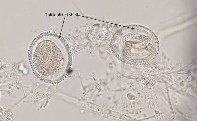
This week's case is a bit unusual in that it is an environmental sample (but the parasite has relevance to human health). The following were seen in a soil sample taken from a child's playground. They are approximately 80 micrometers in greatest dimension. Most likely identification?
Monday, January 25, 2021
Sunday, January 24, 2021
Answer to Case 624
Answer to the Parasite Case of the Week 624: Toxocara sp. eggs. Note that one is fully embryonated and contains an L3 larva. These eggs are found in the feces of the definitive hosts: T. canis in canids and T. cati in felids. Based on the size, the eggs in this case of likely to be those of T. canis, which is slightly small than the eggs of T. cati (80-85 vs 65-75 microns respectively). Of note, these eggs are NOT found in human feces. However, they are a risk to humans if ingested, since eggs with larvae will hatch and can cause visceral larva migrans. That is why finding eggs in the soil of a child's playground is particularly concerning.
The eggs can be identified by their thick outer shell with a pitted surface. It's a very striking appearance.
You can read more about these fascinating zoonotic parasites on the U.S. Centers for Disease Control and Prevention (CDC) DPDx website. Toxocariasis is identified by the CDC as one of 5 Neglected Parasitic Infections in the United States, and is quite prevalent in many places worldwide.
Tuesday, January 19, 2021
Case of the Week 623
The following objects were seen on a peripheral blood film from a patient with chronic, worsening swelling in his groin over the past 5 years. He is from Central Africa. The stain is the Delafield's hematoxylin. Diagnosis?
Sunday, January 17, 2021
Answer to Case 623
Answer to Parasite Case of the Week 623: Wuchereria bancrofti microfilariae
This case showed all of the classic features of W. bancrofti microfilariae, including the sheath which was nicely highlighted by the Delafield's hematoxylin stain. The sheath may not always be seen with routine Giemsa stain; when it is present, it often appears as a negatively-staining outline only. The Delafield's hematoxylin isn't routinely performed in the parasitology laboratory, but it is in all of the classic parasitology texts as an option for highlighting microfilariae sheaths. It's a beautiful stain!
Here are the features of interest:
- Presence of a sheath
- Nuclei do not go all the way to the tip (tip is Without nuclei in Wuchereria)
- Width of the microfilaria is about the same or greater than the surrounding WBCs on thin blood films. This is in contrast to Mansonella spp. which are more slender than the WBC diameter on thin films (important when a sheath is not seen).
Monday, January 11, 2021
Case of the Week 622
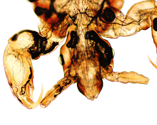
This week's case was graciously donated by Dr. Kyle Rodino, one of our outstanding former Medical Microbiology fellows. The following specimen was submitted to the clinical microbiology laboratory in vodka (which deserves extra points for creativity). Identification?
Sunday, January 10, 2021
Answer to Case 622
Answer to the Parasite Case of the Week 622: drunken Pediculus humanus capitis
There are pretty entertaining and interesting comments that I would encourage you to read if you are interested! Here are some of the key findings in this case:
Thanks again to Dr. Rodino for donating this interesting case.Monday, January 4, 2021
Case of the Week 621
Happy New Year everyone! We are going to kick off the New Year with a fascinating (and challenging) case by Idzi Potters and the Institute of Tropical Medicine, Antwerp.
From Idzi: While I was rummaging through the education-samples, I stumbled upon a strange-looking, small vial, containing a liquid from unknown origin. When I looked at the identification label, I was quite surprised to find a name that sounded like a very exotic parasite…
During a quick microscopical examination, I found eggs of about 60-70 µm in length.
Who can guess which parasite I found? Hint: the source ended up being a cyst near the ear from an African man.
Sunday, January 3, 2021
Answer to Case 621
Answer to the Parasite Case of the Week 621: Poikïlorchis (Achillurbainia) congolensis.
Wow, I am so impressed with how many of you got this identification. This rare parasite was first described in Nature in 1957 in a man from the Belgian Congo.
From Idzi: Poikilorchis congolensis, or alternatively Achillurbainia congolensis -as the genus Poïkilorchis (Fain and Vandepitte, 1957) was regarded by Dollfus as a synonym of Achillurbainia (Dollfus, R. P., 1966. Personal communication).
As far as I have found in the literature, it has been described in humans only eight times up ‘till now, although some authors suggest that some of the reported cases of Paragonimus (especially in Africa) could be in fact cases of Poïkilorchis infection.
Although its hosts are not known for sure, Poïkilorchis congolensis is considered to be a zoonosis, with the common final host probably being leopards (and maybe also giant rats?) and intermediate hosts being probably freshwater crabs.
The infection typically produces subcutaneous retroauricular cysts, which contain as well the eggs as the adults. Nevertheless, in many (human) cases only eggs are found in the cyst.
In the literature, I found human cases in Central and West Africa, Sarawak (Malaysia), possible also one in China…
Idzi and the vial of of Poïkilorchis congolensis eggs.


