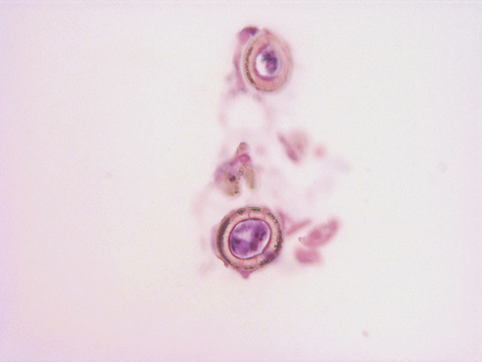
Monday, January 26, 2015

Case of the Week 334

This week's case was generously donated by Dr. Robin Chamberland from Saint Louis University. The specimen is "eyelash scrapings" and the objects shown measure ~180 to 200 microns in length. No patient history is available.


Identification?


Sunday, January 25, 2015

Answer to Case 334

Answer:  Demodex sp. mites

As some of the readers pointed out, the mites are a little beat-up looking (possibly damaged during the scraping process used to obtain the specimen) but you can make out the short legs (arrows below) and elongated abdomen with striations.

These are either D. brevis or D. folliculorum found in hair follicles and sebaceous glands.

Here is a poem from Blaine for this week's case - thanks for using my name in it, Blaine, you're a pal... :)

Living at the base of Bobbi’s right eyebrow
Is a follicle mite, but to know for sure, how?
It’s too slender for scabies or an avian mite
So if you’re ID is Demodex your absolutely right!
Which makes you an entomological maestro, so please take a bow

Monday, January 19, 2015

Case of the Week 333

This week's beautiful case was donated by our loyal reader and contributor, Florida Fan.

It was retrieved from a 70-year-old man and measured nearly 45 cm in length x 0.8 - 1 cm in width. Several ova were expressed and measured 65 x 55 microns. No other clinical history is available.

Identification?


Sunday, January 18, 2015

Answer to Case 333

Answer:  Diphyllobothrium sp. (broad fish tapeworm)

The diagnosis is made by identification of key morphologic characteristics including the broad proglottids with small central uterine structure (arrow) and oval-shaped 65-micron eggs.


Although we can't easily make out the operculum on the egg shown, the abopercular knob (arrow) is visible.

Sunday, January 11, 2015

Case of the Week 332

This week's case was generously donated by Dr. Susan Novak.

A 27 year old woman from Africa had a history of passing worms in her stool since she was a child. After moving to the US, she passed 4 yellow rectangular objects measuring up to 2.0 cm in length x 0.4 cm in width and 0.1 cm in thickness.

These objects were submitted for histologic exam and showed the following:

(H&E, 100x total magnification)

(H&E, 400x total magnification)


 H&E, 1000x total magnification; object measures approximately 35 micrometers in diameter.


Identification?

Saturday, January 10, 2015

Answer to Case 332

Answer:  Taenia sp. proglottids

The 4 structures seen grossly are of the approximately size and shape that would be expected for Taenia proglottids, although they could represent non-parasitic structures as well.  Therefore, additional studies are needed for further characterization of these objects.  In the parasitology laboratory, we always try to examine possible proglottids using a dissecting microscope and look for characteristic structures and eggs.  For suspected Taenia proglottids, we also try to demonstrate the uterine branches so that we can determine the infecting species.

In this case, unfortunately, these proglottids were submitted for histologic sectioning and this limits the degree of identification that can be done somewhat. The presence of calcified stromal bodies called calcareous corpuscles allow us to confirm that these are cestode forms, while the eggs shown in some sections demonstrate a thick outer shell with radial striations which are consistent with Taenia sp.


However, these sections do not show the uterine branches and so the species of Taenia cannot be determined.


Sunday, January 4, 2015

Case of the Week 331

Happy New Year to all of my Readers!  Here is a special case for the start of 2015 - the beginnings of a parasite.  These eggs were found in stool and measure approximately 60 micrometers in greatest dimension.


Identification?
Would you normally expect to find this stage of egg in a human stool specimen?


Saturday, January 3, 2015

Answer to Case 331

Answer:  Ascaris lumbricoides ova, fertile

What makes this case somewhat unique is that there is a larva inside of each egg. As noted by Anon and Dr. Leisure, you would not expect to see an embryonated egg in a fresh stool specimen. Instead, eggs are passed from the human host in the unembryonated form and then develop in the soil into the infectious embryonated form containing a larva.  However, if you have eggs in archived stool specimens - even in specimens preserved in formalin - the eggs can continue to develop as they would in the soil.  That is what happened in this case.  These eggs were seen in a stool specimen that had preserved in 10% formalin for several months, and larvae could be seen moving inside of the eggs!  This is why I always teach my students that formalin will not reliably kill all parasites and that it is important to treat all specimens as if they are potentially infectious.