This week's case was donated by Dr. Lars Westblade. The patient 'coughed' up the following worm (which was still moving!) after approximately 1 month of intermittent hives.
The posterior end was damaged unfortunately, but here is the anterior end:
What is your differential diagnosis?
Monday, February 26, 2018
Sunday, February 25, 2018
Answer to Case 483
Answer: Probable anisakid (Anisakis sp., Pseudoterranova sp., or Contracaeceum sp.)
There was a lot of great discussion on this case! While we can't definitively rule out a migratory immature Ascaris lumbricoides (crawling up from its usual intestinal location), the size of the worm, morphology, and patient history are most consistent with this being an anisakid larva. Anisakiasis occurs in humans following consumption of undercooked fish or seafood containing coiled anisakid larvae. The larvae cannot mature in humans but still have the potential to cause significant problems for their unintended human host. In the 'best case scenario', the larva dies and is passed in stool. If seen by the patient, it may be submitted to the laboratory for identification. A less optimal scenario is what was seen in this case where the live larvae crawls up the esophagus and is 'coughed up' or expelled out of the mouth. While no doubt disturbing, this is still better than the alternative, in which the larva burrows into the gastric or intestinal mucosa, causing significant pain for the host. If the larva is not immediately removed, the patient may experience symptoms for an extended period of time until the larva dies and is absorbed by the host. Rarely, the larva will penetrate the wall of the stomach or intestine and enter the peritoneal cavity, wreaking further havoc. A final, but equally important, complication of exposure to anisakid larvae is development of an allergy to anisakid proteins. This can occur regardless of whether the larva is alive or dead. Sensitized individuals must avoid anisakid-infected fish or risk experiencing serious allergic, or even anaphylactic, reactions, upon re-exposure.
Anisakid larvae can be identified by a few features: they are ~3 cm in length, have 3 fleshy lips just like A. lumbricoides, and also have a very small 'boring' tooth on the anterior end (which can be very difficult to see). Some species also have a posterior spicule called a mucron which is easier to identify. Ascaris doesn't have a boring tooth or posterior mucron, so these are helpful features when seen. Unfortunately the posterior end was damaged during removal so we weren't able to examine it.
What I found to be very interesting about this case was the history of hives, suggesting an allergic reaction to the larva. The time frame of symptoms was also interesting - the patient experienced hives for ~ 1 month before expelling the worm, which indicates that either the larva was present all of that time without causing any gastrointestinal symptoms, or the patient had ongoing exposure to anisakids through his diet. I'd be curious to know - have any of my readers run into a similar case? This is actually the second case I've seen where the patient had been symptomatic for several weeks after presumed exposure and before expelling the larva. This leads me to think that some larva can exist in the host for weeks without burrowing into the gut lining. Please let me know what your experience has been!
Monday, February 19, 2018
Case of the Week 482
This week's case was donated by Dr. Paul Green. The following worm was removed from the eye of a Nigerian man, and was noted to still be moving after removal:
Although it was damaged during removal, the key diagnostic features are still visible.
Anterior end:
Anterior end:
mid-body with disrupted area:
Posterior end:
Identification? What additional specimen could confirm your diagnosis?
Sunday, February 18, 2018
Answer to Case 482
Answer: Loa loa
This great case by Dr. Green shows a gravid female worm with characteristic irregularly-spaced elevations on the cuticle called "bosses". Identification of the bosses allows the adult worm to be differentiated from the similar-appearing adults of many Dirofilaria spp. which have cuticular ridges rather than bosses. William Sears also commented on some of the other characteristic features which aid in identification: a straight buccal canal and muscular esophagus.
Several of you correctly identified the additional test that would be useful for confirming our diagnosis: examination of Giemsa-stained peripheral blood films (collected between 10am and noon) to look for characteristic sheathed microfilariae. This was done, and microfilariae were readily identified:
Note the evidence of a sheath (seen as a negative outline in this case) and nuclei that go to the tip of the tail, consistent with Loa loa microfilariae.
Monday, February 12, 2018
Case of the Week 481
Happy Valentine's Day! Here is a special photo taken by Emily Fernholz, my awesome education specialist. These 2 little worms were found in a concentrated stool specimen and measure approximately 250 micrometers long. Identification?
Sunday, February 11, 2018
Answer to Case 481
Answer: Strongyloides stercoralis rhabditiform larvae
As noted by Florida Fan and Sugar Magnolia, these 2 larvae are "intertwined in romantic interlude" - "performing a romantic ballet". Perfect for Valentine's day!
The identification can easily be made by the size of these nematodes, their short buccal cavities (arrows in image below), and the genital primordium (less easily appreciated here).
Monday, February 5, 2018
Case of the Week 480
Starting us off with the first Monday of February 2018 is another exciting case from Idzi Potters and the Institute of Tropical Medicine, Antwerp.
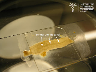
The patient is a 65 year old Spanish woman who brought 3 worm-like structures to her physician. She had expelled the structures 30 minutes prior to presentation, and one was noted to still be moving. She reports no travel outside of Europe.
...and here is the moving one!!
The patient is a 65 year old Spanish woman who brought 3 worm-like structures to her physician. She had expelled the structures 30 minutes prior to presentation, and one was noted to still be moving. She reports no travel outside of Europe.
...and here is the moving one!!
Sunday, February 4, 2018
Answer to Case 480
Answer: Taenia saginata proglottid
As mentioned by Luis, Florida Fan, Atiya, and Sugar Magnolia, these proglottids can be differentiated by other large tapeworms by the size and shape of the proglottids, and the presence of more than 12 uterine branches arising from the central uterine canal/stem:
Remember when counting uterine branches to only count them as they come off the central stem (primary branches), and only on ONE side (not both)!
As some readers mentioned, the proglottids of Taenia saginata are morphologically indistinguishable from those of T. asiatica; however, the patient's lack of travel outside of Europe makes infection with this latter parasite unlikely. Thanks again to Idzi Potters and the Institute of Tropical Medicine, Antwerp, for this interesting case!
Subscribe to:
Posts (Atom)


