Sunday, March 31, 2024

Answer to Case 743

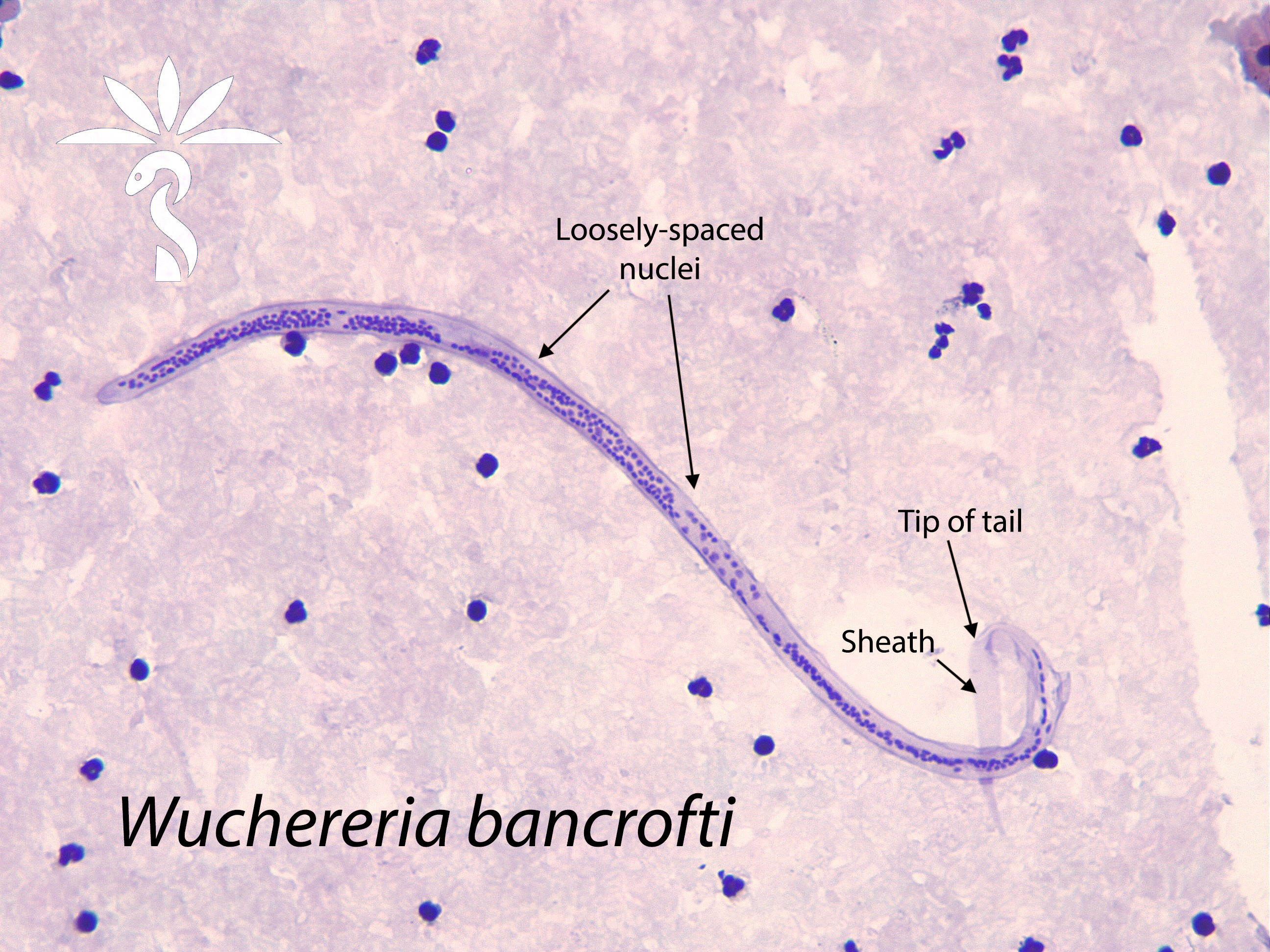
 Answer to the Parasite Case of the Week 743: Brugia timori microfilariae. This was one of the tougher cases, but everyone did a great job narrowing the differential to Brugia. As noted by Florida Fan and Anonymous, we can immediately rule out Mansonella species based on the small size (length and diameter) of the microfilariae. Florida Fan also notes that the sheath is visible, confirming that we are dealing with Loa loa, Wuchereria bancrofti or the Brugia genus. 

He then details his method for coming to an exact diagnosis: "First the column of nuclei is so compact that we can rule out Wuchereria bancrofti, also the nuclei go to the end of the tail. Yet, the two terminal nuclei are distinctly separated from the nuclei column, this rules out any chance of being Loa loa. Now we have the Brugia genus left, but where is the pink sheath pertinent to Brugia malayi when stained with Giemsa stain? As such, we eliminate Brugia malayi. By the elimination process, we’re left with its cousin Brugia timori, the sheath of this one does not stain pink with Giemsa stain. Now let’s check the map of the lesser Sundae island, the Alor island is right North of Timor."

I will also point out that the head space is longer than that of B. malayi and there are a larger number of single-file nuclei going towards the tail. These features are more subtle and can be difficult to appreciate. I've done my best to point out some of them in the image below.

Thank you all for sticking with us on our microfilariae journey. The microfilariae can be very challenging to identify, and are rare in many parts of the world, so our lab staff may not have the opportunity to examine these organisms outside of their EQA/PT schema. 


Monday, March 25, 2024

Case of the Week 742

This week's case features another beautiful example of microfilariae in blood. The patient is from sub-Saharan Africa and presents with chronic swelling of his left leg. The microfilariae measure approximately 270 ยตm in length. 

Carazzi stain (Knott's concentration):


Giemsa Stain (thick blood film)

What is your identification? What time should blood be collected for this examination?

Sunday, March 24, 2024

Answer to Case 742

 Answer to the Parasite Case of the Week 742: Wuchereria bancrofti microfilariae.

I really enjoyed reading the comments on this case. FloridaFan provided this excellent description of his approach to microfilariae identification: "First the width of the worm is about the same as that of the surrounding neutrophils. Second, its length is greater than 200 micrometers. Third, it has a sheath. Now we know we are dealing with Loa loa, Wuchereria, or the Brugia ones, not with any little pesky Brugia. The next consideration is that the column of nuclei is continuous, the terminal nuclei are not separate from the immediate anterior nuclei. This eliminates the possibility of the Brugia malayi and timori, over that these are “far fetched” geographically. We are left with two candidates Loa loa and Wuchereria bancrofti. Though the tail is not so obvious, the Carrazi stain did show that the nuclei column terminates well short from the end of the tail. This rules out Loa loa. The only culprit left is Wuchereria bancrofti.

An anonymous commenter also noted that "Its nuclear column is relatively loose, and individual nuclei can be visualized throughout the column. The sheath is does not stain pink with Giemsa stain (as in Brugia malayi)."

These features can be nicely visualized in the photo of the Carazzi-stained Knott's concentration. Note that the Carazzi stain is a hematoxylin-based stain (rather than conventional Giemsa) that helps to demonstrate the sheath. It is a very useful stain to have!

Thanks again to Idzi Potters and the Institute of Tropical Medicine, Anwerp, for this beautiful case.


Monday, March 18, 2024

Case of the Week 741

Wow, we are already on our 3rd filarial case! The following lovely case from Idzi Potters and the Institute of Tropical Medicine, Antwerp, is another microfilaria found in blood. 

The patient is a middle aged male farmer from Central America who was noted to have mild eosinophilia on routine complete blood count. He is otherwise asymptomatic. The microfilariae measure approximately 175 micrometers in length. What is your identification?


Sunday, March 17, 2024

Answer to Case 741

 Answer to the Parasite Case of the Week 741: Mansonella ozzardi

As many of you noted, we can get to an identification of Mansonella sp. by the small size (length of just 175 micrometers, and the width less than the surrounding white blood cells). There is also no visible sheath, which is supportive of the diagnosis. 

So the next question is - which Mansonella is present?? As FloridaFan mentioned, the tip of the tail is pointed rather than blunt, which leads us away from M. perstans. Also, the source is not skin snips, so M. streptocerca is unlikely. That leaves M. ozzardi by default. 

To confirm this, we would look at the tail to see if the nuclei go to the tip (M. perstans) or not (M. ozzardi). It's a bit hard to tell in this case, but if you look closely, you can see that the nuclei DON'T go to the tip of the tail. So - Mansonella ozzardi, it is!

Congratulations to those of you who were able to get to Mansonella species. The microfilariae can be hard to identify, so this is definitely advanced  parasitology!

Monday, March 11, 2024

Case of the Week 740

Welcome to our next filarial case by Idzi Potters and the Institute of Tropical Medicine, Antwerp. This week features the following lovely microfilariae seen in a Giemsa-stained thick blood film. They measure approximately 220 micrometers in length. Identification?


Sunday, March 10, 2024

Answer to Case 740

Answer to the Parasite Case of the Week 741: Brugia malayi 

Thanks to everyone who wrote in. This is one of my favorite microfilariae! 

This case had 2 classic features that facilitated the identification: the pink sheath and separation of the 2 terminal nuclei in the tail (see arrows below). 

As I noted in the case last week, the sheath isn't always seen. Therefore, the larger length and tail nuclei configuration can allow for the identification, even when the sheath is absent. 

Sunday, March 3, 2024

It's Finally Here - Filariasis Month with Idzi Potters! Case of the Week 739

Dear Readers,

Welcome to Filariasis Month! We will actually have TWO months of filariae for you as there are so many to cover and so many beautiful cases by Idzi and the Institute of Tropical Medicine, Antwerp.

Idzi and I thought that we should start out with an 'easy' one. The following objects were seen on a blood smear from a patient living in Gabon. They are approximately 250 micrometers long. What is your identification? Bonus question: what additional laboratory test is important for guiding treatment?


Giemsa stained blood films:


Carazzi stain, Knott's concentration:


Saturday, March 2, 2024

Answer to Case 739

 Answer to Parasite Case of the Week 739: Loa loa microfilariae

Thanks to everyone who wrote in with comments. We received a lot of different responses including some of the sheathed and unsheathed microfilariae. Therefore, this is a great time to review my approach to identifying microfilariae in blood specimens. You can also read this article I wrote with Blaine Mathison and Marc Couturier that provides a diagnostic algorithm for microfilariae in blood. In this algorithm, we recommend first measuring the length of the microfilariae. If they are small (<200 micrometers long), then it is likely to be one of the Mansonella species. Mansonella are not sheathed, small, and quite narrow. Their width is smaller than the diameter of a neutrophil or eosinophil. If, on the other hand, the microfilariae are relatively large (>200 micrometers), then you are dealing with Wuchereria bancrofti, Loa loa, or one of the Brugia species. These are sheathed microfilariae, but the sheath may not always be readily visible on Giemsa stain. Therefore, the size is a more reliable diagnostic feature. To better visualize the sheath, a hematoxylin stain can be performed such as the Carazzi or Delafield hematoxylin stain. (Note: the hematoxylin and eosin stain used in histopathology preparations can also be used, so if you don't have a hematoxylin stain for microfilariae, you can ask for help from your friends in anatomic pathology!)  These larger microfilariae can then be differentiated by characteristics of their head and tail spaces. 

With that introduction, let's turn to the specifics of this week's case. I forgot to provide the length - my apologies! - I've now added it to the case description (~250 micrometers long). You can also get a general sense of the size of the microfilariae by comparing their diameter to the surrounding white blood cells. Note in this image below that the width of sheathed microfilariae such as Loa loa is slightly greater than the diameter of the neighboring eosinophils. Also, there is a hint of sheath (arrows). In contrast, the Mansonella perstans microfilaria in the image on the right is narrower than the surrounding eosinophils. 

Importantly, you can also see a sheath with the Carazzi stain that Idzi provided. Therefore, we can rule out the two Mansonella species found in blood by this finding alone. 

Note that the nuclei go to the tip of the tail, which makes this a classic case of Loa loa. (one of my favorite memory aids is that the nuclei 'flow-a flow-a to the tip in Loa loa). 


To answer my second question - the additional study that should be considered in this case is calculation of the microfilarial burden since patients with >8,000 microfilariae/mL are at risk of encephalopathy when treated with diethylcarbamazine (DEC).

Thanks again to Idzi Potters and the Institute of Tropical Medicine, Antwerp, for this great case! The next case will come out early next week.