Monday, December 28, 2015

Case of the Week 378

This week's case was donated from one of our superstar pathology residents, Dr. Heidi Lehrke. The specimen was a tan-white tissue, approximately 4 mm x 1 mm x 1 mm, that was obtained during colonoscopy and submitted for histopathologic processing. Stain shown is H&E. Identification?

40x total magnification

100x total magnification

400x total magnification

Sunday, December 27, 2015

Answer to Case 378

Answer: Dipylidium caninum

As mentioned by Florida Fan, the key to identifying this case is to recognize the thin outer cuticle (consistent with a worm) and the clusters of eggs within thin-shelled packets.

 On higher magnification, the negative outline of hooklets can be seen within some of the eggs (if you use your imagination a bit).

And now - a poem from Blaine:

‘Waiter!’ the man shouted, ‘there’s a fly in my soup’
For which he was corrected, ‘No that’s a flea in the Siphonaptera group!’
‘What does it matter, it’s still a darn bug!!!’
To which replied our waiter who is entomologically smug:
‘But if you eat that lil’ critter there, you’ll have D. caninum in your poop!'

Monday, December 21, 2015

Case of the Week 377

Happy Holidays to all of my Creepy Dreadful Wonderful Parasite blog readers!

Here is a fun holiday-themed case: 'Santa' delivering 'gifts.' Who is Santa, and what is in the gifts?


Sunday, December 20, 2015

Answer to Case 377

Answer:  Pthirus pubis, the crab louse; also nicknamed here 'Santa' louse.

As so nicely described by Kamran and Florida Fan, 'Santa' louse is coming down the hair shaft to leave a gift of a nit glued onto a hair shaft!

And a poem from Blaine:
Nice guys and gals will get toys in their stocking
But the naughty ones are sure to get something more shocking!
For the gift that keeps on giving is a parasitic louse
And that’s what you get for stepping out on your spouse!
So you’d better be good ‘cuz Santa Crab’s always watching!

Happy Holidays to all of my Readers! May the parasites you see not be your own.

Monday, December 14, 2015

Case of the Week 376

This week's case includes beautiful (and creepy) photographs and a movie from Dr. Graham Hickling at the University of Tennessee.

The following egg-laying arthropod was observed last month in her natural forest habitat. Identification?


Once you look at the photos, take a look at this video (set to music). It gives part of the answer away, but still leaves you with the task of determining the genus of this organism:
https://vimeo.com/148692758

Enjoy!

Sunday, December 13, 2015

Answer to Case 376

Answer: Ixodes sp. adult female tick, laying eggs.

If you watched the video, then you probably noticed the "inverted U-shaped" anal groove (arrow below), which allows us to identify this tick as an Ixodes species.


So for those of you wondering what it looks like when all of these eggs hatch, I have some amazing follow-up photos for you from Dr. Hickling:


Tuesday, December 8, 2015

Case of the Week 375

This week's case is a bit of a challenge, and not something that has been featured on this blog before. It was generously donated by Dr. Kamran Kadkhoda.

The following objects were seen in the stool from a 30-year-old woman from Canada. No travel history is available. They measure approximately 50 micrometers in length. Identification?


Monday, December 7, 2015

Answer to Case 375

Answer: Not a human parasite; most consistent with a member of the Myxozoa, aquatic parasitic animals.

These are an incidental finding in human stool specimens, and usually indicate that the individual has recently consumed fish such as salmon. Arthur points out that microbiologists should be aware of this finding because of the very real possibility of mistaking these structures as human spermatozoa. This is especially important when dealing with cases of children and suspected sexual abuse. Here is the article that he recommended:

McClelland, R. S., D. M. Murphy, and D. K. Cone. "Report of Spores of Henneguya Salminicola (Myxozoa) in Human Stool Specimens: Possible Source of Confusion with Human Spermatozoa." Journal of Clinical Microbiology 35.11 (1997): 2815-818.

You can differentiate these structures from human spermatozoa by the consistent presence of 2 tail processes (rather than 1 tail in spermatozoa; only occasionally are 2 tails present), larger head structure with 2 polar capsules, and longer tail in relation to the head. Spermatozoa also have a longer defined neck region. Unfortunately, the appearances can be very similar, and therefore getting a dietary history may also be helpful.

And now a fun poem from Blaine!

Looking at this case after the last, you might ask ‘What is Bobbi doing?’
Pubic lice followed by spermatozoa, does she think this is amusing?
Alas this is only Myxozoa spuriously passed in the stool
After eating parasitized fish caught in a contaminated pool
And so Bobbi can now be absolved of a case that some might be misconstruing

Sunday, November 29, 2015

Case of the Week 374

This lovely little family was found on a 20 year old male. Thanks to Florida Fan for donating this case!  Identification?


Saturday, November 28, 2015

Answer to Case 374

Answer: Pthirus pubis ('crab' lice)

As noted by Arthur, "two females are apparent by the presence of a bifurcate posterior, whereas the male has a rounded posterior."

Note that the claws on first pair of legs are slender, compared to the larger claws on the 2nd and 3rd pairs of legs.
To answer MicrOlivier Biologist's question about this species being endangered due to modern practices of pubic hair removal, I have to say to say that it still seems to turn up with some regularity in the routine clinical microbiology lab, so clearly there are a population of 'adequately' hairy individuals out there!

Thanks again to Florida Fan for donating this case, and also to all of my readers for their excellent comments and questions.

Monday, November 23, 2015

Case of the Week 373

The following structures were found in stool.  They measure approximately 60 micrometers in greatest dimension. Identification?


Sunday, November 22, 2015

Answer to Case 373

Answer: pinworm eggs - with a newly emerging larva. Kudos to Felicity in my lab for catching this worm in action!

Note the range of eggs seen here, including a classic oval/elliptical eggs with flattened side, an egg containing a larva, and lastly, the emerging larva.

And here's a poem from Blaine!

So many nematodes in the gut, it’s hard to tell which is which
But Enterobius vermicularis fulfills a particular niche
For she likes to come out at night
And oviposit at the site
That is sure to make your booty itch!

Sunday, November 15, 2015

Case of the Week 372

Several objects were removed from a foot wound of a 70 year old woman. Representative photos of one of these objects is shown below. Identification?


Saturday, November 14, 2015

Answer to Case 372

Answer: Musca domestica (common housefly) larva

Unlike obligate parasites such as Dermatobia hominis, Musca domestica is a cause of accidental myiasis. While M. domestica larvae are usually found in nutrient rich organic material such as compost and manure, they can occasionally infest human tissues. The adult flies can also serve as a mechanical vector for infectious agents including bacteria, viruses and parasites.

The CDC has an excellent online reference for identifying medically important flies that you can access HERE. As you can see from the identification key, fly larvae are identified by a number of features including their overall shape and characteristics of their posterior spiracles. The line drawing of the M. domestica spiracles is shown below, next to the photograph from this case for this comparison.


Tuesday, November 10, 2015

Case of the Week 371

This week's case was generously donated by Dr. Zhenwen Zhou, MD, from the Department of Laboratory Medicine at Guangzhou Women and Children's Medical Center, Guangzhou Medical College, in Guangzhou, People's Republic of China.

The patient is a middle-aged man who found the object below in his stool. He reports eating rare beef approximately 2 months earlier.


Identification?

Sunday, November 8, 2015

Answer to Case 371

Answer: Taenia species tapeworm segment.

The segment shown is notable for the fact that it is still moving (!) as well as the fact that the proglottids are longer than they are wide. The latter feature, in addition to the large size of the worm, supports an identification of Taenia species (as compared to Diphyllobothrium spp., Dipylidium caninum, and Hymenolepis spp. Unfortunately, we are not given any additional morphologic features that would allow us to determine which Taenia species is shown (such as the uterine branching pattern or scolex). However, Florida Fan and other readers correctly note, that the history of eating undercooked beef supports the supposition that this is T. saginata. In comparison, Taenia solium and Taenia asciatica are acquired by ingesting undercooked pig (muscle, or in the case of T. asciatica, liver and possibly other organs).

Sunday, November 1, 2015

Case of the Week 370

Happy Halloween dear readers!
Like last year, this week's case highlights some of the costumes and goodies from my annual Halloween Party. 

First - the costumes! Here are the amebae ladies. Can you guess which is which? I'll give you a hint in that they are all morphologically identical, but one is slightly smaller than the others.
Here are 2 of our Clinical Microbiology Fellows - the "FakeArray" and the baker (scroll down further to see photos of her goodies) 

Close up of the "FakeArray" Sugar Overload Panel, which detects 24 sweet targets. (The actual FilmArray(R) detects multiple enteric pathogens, including 4 parasites)

And here is the party's hostess, parasite gal in a Peromyscus leucopus costume. Note my Ixodes scapularis accessories.


And last, but not least, the gory baker's cupcakes. Can you tell which parasite is shown on the cupcake with the pretzel stick?


Saturday, October 31, 2015

Answer to Case 370

Answer: Lots of Halloween goodies.

As Arthur notes, the Entamoeba ladies include Entamoeba histolytica and E. dispar. There are also E. moshkovskii and E. bangladeshi - all morphologically indistinguishable of course! The smaller Entamoeba was E. hartmanni.  

The cupcakes were amazing - all courtesy of our Clinical Microbiology fellow, Dr. Rachael Liesman. As Yasir and Florida Fan mention, the cupcake with the pretzel stick represents Dracunculus medinensis, the Guinea worm, being pulled from a wound and wrapped around a stick. The equally clever cupcake right above this is a wound full of maggots. Yum. (they were actually delicious cupcakes).


Thanks for all of the great comments.

Sunday, October 25, 2015

Case of the Week 369

This week's case was generously donated by Dr. Jane Hata. Several motile structures were seen in a bronchoalveolar lavage from an elderly woman with rapidly progressing shortness of breath and edema. She had a history of chronic heart failure and was receiving immunosuppressive medications for a renal transplant some years prior.

Identification?


Saturday, October 24, 2015

Answer to Case 369

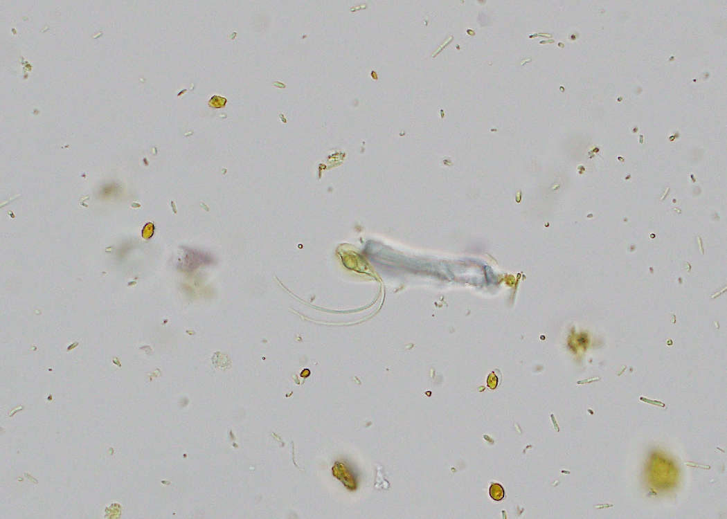
Answer:  ciliated epithelial cells and detached ciliary tufts (ciliocytophthoria)

This case demonstrates a convincing parasite mimic that is commonly seen in deep respiratory specimens: detached and intact cilia from respiratory epithelial host cells. The cilia on these structures will remain motile long after the cells are sloughed from the human host and thus resemble ciliated or flagellate parasites such as Balantidium coli and Trichomonas tenax. However, the morphologic features allow for clear differentiation of these objects. Detached ciliary tufts (DCTs) are relatively small (5-15 micrometers in diameter), the cilia are present in a dense mat attached to a terminal plate, and the cilia beat in a rhythmic motion without propelling the object forward.

In comparison, Arthur Morris notes that "B. coli is very (very!) large in its ciliated trophozoite form" (40-200 micrometers) "and is covered in short cilia rather than a localized patch of long cilia" (as seen in this case). "It also has a distinct peristome and kidney-shaped macronucleus which is not present here." The cilia on B. coli cause it to move in a 'rotary' or 'boring' motion.

Similarly, Trichomonas tenax is easily differentiated from DCTs by morphology. It has only 4 apical flagellae (rather than a tuft of dense cilia) and demonstrates a 'jerky' motility pattern.

Of note, the free-living amebae that infect humans (e.g. Naegleria fowleri) do not have a flagellate form in the human body and therefore should not enter the differential diagnosis when DCTs are seen. Also, Lophomonas blattarum is a parasite of cockroaches and has not been convincingly described from humans; instead, it has been misdiagnosed in cases where DCTs were observed.

HERE is another case from my blog, showing Papanicolaou-stained DCTs.

Also, HERE is an article (in Chinese) that discusses how previously-reported cases of L. blattarum infection were actually cases of misidentified bronchial ciliated epithelial cells.


Tuesday, October 20, 2015

Case of the Week 368

The following structures were seen in a Papanicolaou-stained sputum specimen from a patient with chronic obstructive pulmonary disease. Identification?


Sunday, October 18, 2015

Answer to Case 368

Answer:  Not a parasite; Curschmann spirals

As nicely described by Anon, Curschmann spirals are spiral-shaped mucous plugs that are commonly seen in patients with asthma and other chronic obstructive lung diseases. They are formed when thick mucous accumulates in the patient's small airways and forms a plug or cast of the airways. These casts can then released into lower respiratory specimens such as sputum and bronchoalveolar lavage (BAL) fluid and be seen on cytology and microbiology specimens. Even though they can superficially resemble worms, they are easily differentiated by their lack of internal structures, long irregular shape, and variable diameter.


Monday, October 12, 2015

Case of the Week 367

This week's case is a Giemsa-stained sputum specimen from a patient with abrupt onset of productive cough and expectoration of "salty-tasting" fluid. Images are shown at 400. and 1000x. Identification?


Sunday, October 11, 2015

Answer to Case 367

Answer:  Echinococcus sp.

The main diagnostic features of this case are the characteristic protoscoleces with suckers and hooklets in a background of free calcified bodies (calcareous corpuscles).  Free hooklets are also present. The protoscoleces, calcareous corpuscles and free hooklets make up the material that is commonly referred to as "hydatid sand". Calcareous corpuscles are also seen in the stroma of each protoscolex. These calcified bodies are not unique to Echinococcus spp., but are found in all cestodes (larva and adults)


As many readers mentioned, this is most likely to be echinococcosis due to Echinococcus granulosus, since this is the most common species to infect humans. Yasir correctly notes that E. multilocularis rarely infects humans and almost exclusively involves the liver rather than the lungs.

Dima also notes that this could also be E. vogeli or E. oligarthrus, although these are both very rare in humans. Imaging studies would help differentiate cystic echinococcosis due to E. granulosus from alveolar or polycystic echinococcosis due to E. multilocularis and E.vogeli respectively.