Monday, April 26, 2021

Case of the Week 636

This week's case was donated by one of our intrepid infectious diseases fellows, Dr. Geno Tai, who was travelling across country and found these arthropods in his lodging. Identification? Photos and video courtesy of his friend and travel-mate, Dr. Chris Hwang.


Sunday, April 25, 2021

Answer to Case 636

 Answer to the Parasite Case of the Week 636: Cimex sp. 

Given the location (hotel in North America), this is most likely Cimex lectularis, the common bed bug.  However, we would want to have a closer examination to be sure. It looks like we have a couple of different life cycle stages here. Also, at least one squished well-fed one - nice job Geno!  


Here is a good resource for information.

Monday, April 19, 2021

Case of the Week 635

This week's case features a fun finding in a concentrated wet prep of stool stained with iodine. The case and photograph are from Blaine Mathison and Madison Sant. The object below measures approximately 35 micrometers in diameter. Identification?


Sunday, April 18, 2021

Answer to Case 635

 Answer: Entamoeba coli, supernucleate cyst. Typical E. coli cysts have 8 nuclei, but I'm counting at least 18 nuclei in this one. My first thought when I saw this photograph was - holy moly E. coli! Thanks again to Blaine and Madison for donating this fun case.


Monday, April 12, 2021

Case of the Week 634

 This week's case was generously donated by Dr. José Poloni. The following was found in a stool specimen. Identification? Significance? 

(you may need to click twice on the video below to play)


Sunday, April 11, 2021

Answer to Case 634

 Answer to Parasite Case of the Week 634: Free-living mite, likely contaminant during specimen collection/preparation.

Note the 8 legs (consistent with a member of the arachnidae) and the long spines (setae). It is easier to appreciate all 8 legs by carefully watching the video in this case.


As noted by Marc Couturier, "I mite have to say this is a contamination of the preparation and not coming from stool."  I agree with this assessment! While this mite could have been ingested from a food source (e.g., mimolette cheese; see my previous post), I find it highly unlikely that the mite would still be alive and active after passing through the intestinal tract. I also find the presence of squamous epithelial cells nearby to be rather suspicious for environmental contamination. 

Thanks again to Dr. José Poloni for donating this interesting case, and to all of the readers who wrote in with their helpful commentary. 


Monday, April 5, 2021

Case of the Week 633

 This week's case was generously donated by Dr. Neil Anderson. The following structure was retrieved from the common bile duct during endoscopic retrograde cholangiopancreatography (ERCP). The patient is a refugee from Tanzania who presented with intermittent abdominal pain, distended gallbladder and hepatosplenomegaly. This was one of many "worm like" structures noted on ERCP.  Unfortunately this object appeared to tear during removal. The portion submitted measures several centimeters in length. No identifying external structures were identified.


Dr. Anderson's team tried to express eggs from this structure but was unsuccessful. In an effort to learn more about the nature of this object, a portion of it was submitted for histopathologic processing. Representative sections revealed the following: 


What is the most likely identification?


Sunday, April 4, 2021

Answer to Case 633

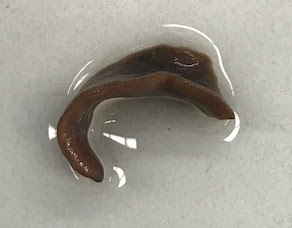
 Answer to Parasite Case of the Week 633: Fasciola hepatica 

Although the fluke was sadly torn in half during retrieval, it has all of the features that allows us to identify it:

As a trematode - it has the flat, leaf-like body shape of a platyhelminth belonging to the Trematoda phylum. On histopathologic examination, trematodes have an outer tegument (with microvillus border, and often with spines), spongy parenchyma with no large cavities, and a digestive tract. Cestodes have a similar appearance, but may have a large cavity (depending on the species and stage), do NOT have a digestive tract or tegumental spines, and have calcareous corpuscles in the stroma.  


I don't believe I see see ovarian tubules in this section, although it is hard to tell from this image alone.  I also don't see any eggs. This would make sense as Dr. Anderson was not able to express any eggs from the portion of the fluke that he received in the laboratory.

As Fasciola hepatica specifically - the large size of this structure allows us to identify it as either Fasciola hepatica or F. gigantica. We can further identify this fluke as F. hepatica based on the presence of pointed tegumental spines. F. gigantica, in comparison, has tegumental spines with blunt/flattened ends. Other morphologic features (e.g., overall size of the adult and its eggs, features of the acetabulum) can also be used to differentiate F. hepatica and F. gigantica.


Thanks again to Dr. Anderson for sharing this case with us!