This week's case is by Idzi Potters and the Institute of Tropical Medicine Antwerp. As always, he has an interesting challenge for us! the following were seen in a wet preparation of the stool concentrate. Identification?


This week's case is by Idzi Potters and the Institute of Tropical Medicine Antwerp. As always, he has an interesting challenge for us! the following were seen in a wet preparation of the stool concentrate. Identification?
Answer to the Parasite Case of the Week 671: Infertile Ascaris lumbricoides eggs, many decorticated.
Decorticated eggs can pose a diagnostic challenge since they resemble eggs of other helminths such as Ancylostoma duodenale, Necator americanus, Trichostrongylus species, Fasciola hepatica, and Fasciolopsis buski,. However, they can usually be differentiated by their thicker wall, size, lack of operculum (seen with the fasciolids), and other characteristic features.

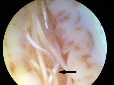
This week's case was generously donated by Dr. Steven O'Connor. He noted multiple worms in an resected appendix, and captured the following images using a dissecting microscope. Identification?
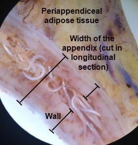
Answer to the Parasite Case of the Week 670: Enterobius vermicularis (pinworm) adults within the lumen of the appendix. This is a classic ectopic location for pinworm. While there is some evidence that they may cause appendicitis in this setting, it could just be an incidental coincidence, given how common both appendicitis and pinworm infection are!
For those of you not familiar with the gross appearance of the appendix, I've included an annotated version below for clarification. You can also compare the size of the appendix to the rest of the intestinal tract from this Mayo Clinic image.
The small size of the appendix will allow you to rule out Ascaris lumbricoides here, since the adult worms would be about the same size as the inside of the appendix. Having said that, Ascaris will occasionally migrate into the appendix and possibly lead to appendicitis, so it's an important consideration!
The other worm to consider in this case is Trichuris trichiura (whipworm), would can also occasionally be seen within the appendix. While it's hard to rule out whipworm in this case, there are occasional worms in which a narrow 'pin-shaped' tail can be seen, which is characteristic of female pinworms.
Whipworms have a broad, rounded tail in comparison.
Here's another fun parasite histopathology case for you: a full-thickness section of bladder wall from an Egyptian man with invasive bladder cancer (not shown here):

Diagnosis?
Answer to the Parasite Case of the Week 669: Schistosoma haematobium adults and eggs - a classic case!
As noted by Idzi, "Looking at site of infection and geography, most probably S. haematobium. Section shows adult fluke and numerous non-calcified viable eggs (because I can see the cephalic gland, surrounded by nerve cells - a beautiful bullseye). In some eggs I seem to discern a small terminal spine, which supports S. haematobium."
This is a great example, given that we rarely see the adults in tissue. Here are some of the key findings:


This week's case is a lovely cross-section of an arthropod embedded within the epidermis. The patient is a middle-aged woman with a lesion on her foot after returning from the Caribbean on holiday. Here is a still image of the digital slide:

Identification?
Answer to the Parasite Case of the Week 668: Tunga penetrans (likely one adult female flea) embedded in skin. Hopefully you all had a chance to look at the digital slide HERE. It is fun to zoom around on the slide and see the various features.
The follow photos show some of the key diagnostic features:


Happy New Year! Here's a great case to start off 2022. A large structure (~25 cm-long) submitted to the Clinical Parasitology lab (found in stool). Identification?

Photo is courtesy of Heather Morris.
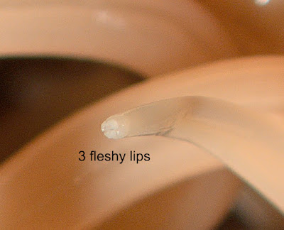
Answer to the Parasite Case of the Week 667: Ascaris lumbricoides.
This adult nematode is easy to identify when found in human stool, or expelled through the mouth, nose, or anus, due to its large size and characteristic 3 fleshy lips. Importantly, anisakid larvae also have 3 fleshy lips, and must therefore be differentiated from immature Ascaris when expelled from the human gastrointestinal tract. This can be accomplished by examining the characteristics of the intestinal tract, mouth and tail.