Friday, December 23, 2011

Case of the Week 189

Dear Readership of Creepy Dreadful Wonderful Parasites,
May you have a wonderful holiday season with friends and family! Thank you for your continued support and comments.

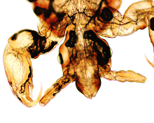
Here is the 'case' for this week - a simple identification:

Thursday, December 22, 2011

Answer to Case 189

Answer: Phthirus pubis, a.k.a the crab louse, or as my readers said:
"the Yuletide pubic louse", "Pthirus barbae viridis: green bearded louse", "Pine-scented pubic louse", and "Louse found in Bad Santa's trousers"

Happy 2012!
Parasite gal

Monday, December 12, 2011

Case of the Week 188

The following objects were found in the clothing of an 18 month old child. They measure approximately 3mm in length.


By manipulating them with a wooden applicator stick, the following was expressed out of these objects (CLICK ON IMAGES TO ENLARGE):

(100x original magnification)


(400x original magnification)


Thanks to Emily in my lab for these beautiful photos!

Sunday, December 11, 2011

Answer to Case 188

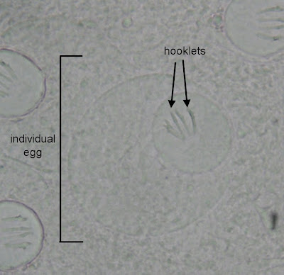
Answer: Proglottids and egg packets of Dipylidium caninum, the double-pored dog tape worm.

This small tape worm is acquired through ingestion of infected Ctenocephalides spp. fleas, prompting one of my readers to ask if the child got infected by ingesting the specimen from the previous case of the week! Another reader, MicrobeMan, had a similar inquiry "Was it merely coincidental that you posted Ctenocephalides last week? Regardless, it's very appropriate, seeing as that both adult C. canis and C. felis can carry the cysticercoid form of D. caninum to people."

To answer these questions - it was actually a complete coincidence that we happened to have a Ctenocephalides flea and D. caninum proglottids submitted to my lab in such a short time period of time, and I wasn't really considering the coincidence when posting these 2 cases. So good pick up for those of you who noticed!

I like this case because it has several classic features which allow for definitive identification. The small "rice grain" sized and shaped proglottids are classic, and the egg packets that were expressed from the proglottids are diagnostic, despite being immature. Note that the material surrounding the eggs is very granular, which is something I've seen frequently when eggs are prematurely expressed from the proglottids, rather than being released naturally. So don't let that deter you from making a diagnosis if the other characteristic features such as egg packets and hooklets inside of egg-like structures are present

Monday, December 5, 2011

Case of the Week 187

Since I am at the ASTMH annual meeting at the moment, I will keep this case short and sweet:

The following was submitted for identification to the clinical parasitology laboratory. It was difficult to get it to hold still, since it was jumping between lids of a closed petri dish. It measures approximately 2 mm in length. (CLICK ON BOTTOM RIGHT OF MOVIE TO ENLARGE)

Identification?

Sunday, December 4, 2011

Answer to Case 187

Answer: Flea

Everyone who wrote in got this correct, despite the fact that the flea wasn't really staying still. We eventually took Blaine's advice and killed it (by placing it in ethanol). We then mounted it on a slide and got a much clearer picture of the flea, showing the genal and pronotal combs that are characteristic for Ctenocephalides species:


The dog and cat fleas (Ctenocephalides canis and C. felis respectively) can be further differentiated based on the shape of their head and "tooth" length of the genal comb.

Monday, November 28, 2011

Case of the Week 186

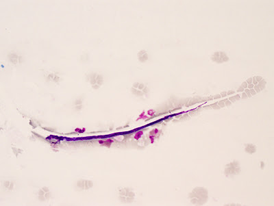
The following was an incidental finding on pap smear. Identification? (CLICK ON IMAGES TO ENLARGE)

Pap stain, 400x original magnification


Pap stain, 1000x original magnification

Sunday, November 27, 2011

Answer to Case 186

Answer: Not a human parasite

I apologize to my readers that I don't know exactly WHAT this is. A consensus of knowledgeable parasitologists thought that this was not a human parasite, and possibly an artifact or fungal element. Meanwhile, my mycology colleagues did NOT think that this was a fungal element. Responses from the viewers also included Turbatrix aceti (vinegar eel) and hair.

The good news for this patient is that this is not a human pathogen and simply an incidental finding.

Thank you all for writing in with your interesting ideas!

Friday, November 18, 2011

Case of the Week 185

The following was worm was seen emerging from a cricket:


Here are photos of the worm itself:


Identification?
Many thanks to Jason Pelletier who took the first photograph and Heidi Pierce who shared this cool case with me!

Thursday, November 17, 2011

Answer to Case 185

Answer: Horsehair worm, also known as the Gordian worm, in the group Nematomorpha.

Over 300 species in this group have been described. They are parasitic in their larval stage for various insects and crustaceans, and emerge from the host (as seen in this case) as free-living adults. The adults are typically found near or in water and are known to twist themselves into complex knots (resembling Gordian knots).

These worms superficially resemble nematodes and they are not uncommonly submitted to our lab for identification; however, they are not parasites of humans or other mammals.

Monday, November 14, 2011

Case of the Week 184

The following images are from a Papanicolaou-stained bronchoalveolar lavage obtained from a 70 year old man with increasing respiratory distress. His medical history is significant for hemodialysis-dependent end-stage renal disease and vasculitis (treated with cytoxan).

Diagnosis? (CLICK ON IMAGES TO ENLARGE)
Any thoughts on what the last image represents?

x400 original magnification


x600 original magnification


Many thanks to Dr. Steven Goodman for donating this case!

Sunday, November 13, 2011

Answer to Case 184

Answer: Probable Strongyloides stercoralis larvae

Higher magnification would be needed to confirm the characteristic features of S. stercoralis filariform larvae (the stage that migrates to the lung during autoinfection). However, the overall appearance and clinical presentation is most consistent with strongyloidiasis hyperinfection.

To answer my question about the last photo - this does not represent a parasite, but might represent a fiber with proteinaceous debris. I was curious to see what others had to say, but no one commented.

Thanks to everyone for writing in!

Monday, November 7, 2011

Case of the Week 183

The following were seen on Giemsa-stained peripheral blood films. The objects shown measure approximately 220 microns in length. Identification? (CLICK ON IMAGES TO ENLARGE)

x1000 original magnification


Sunday, November 6, 2011

Answer to Case 183

Answer: microfilaria of Brugia malayi

The diagnostic features of this case include the brightly staining pink sheath with routine Giemsa staining, relatively small size, and both terminal and subterminal nuclei (see below; click on image to enlarge).

Monday, October 31, 2011

Case of the Week 182

For this week's case, I thought you might enjoy seeing a 'potpourri of parasites' from a stool concentrate wet preparation.

Which eggs are shown here?

Sunday, October 30, 2011

Answer to Case 182

Answer: Ova of Ascaris lumbricoides (upper center/right), Trichuris trichiura (left), and Taenia species (bottom, center/right). The eggs of Taenia solium and T. saginata cannot be differentiated by morphology, so Taenia species is an appropriate result.

Thank you for all of the responses on this case. They were all correct!

Monday, October 24, 2011

Case of the Week 181

The following were incidental findings on seen on intestinal biopsies. Diagnosis? (CLICK ON IMAGES TO ENLARGE)
x20 original magnification


x40 original magnification


x100 original magnification


x200 original magnification


x400 original magnification

Sunday, October 23, 2011

Answer to Case 181

Answer: Whipworm (Trichuris trichiura)

Shown in this case are sections of a round worm in the colonic mucosa. The presence of cross-sections within the mucosa raise the possibility of several nematodes, including Strongyloides stercoralis, Capillaria philippinensis, and Trichuris trichiura, with the latter being supported by the presence of free-hanging sections of worm in the intestinal lumen (recall that the smaller head of Trichuris embeds itself into the colonic mucosa (arrows below), leaving the larger body hanging free in the lumen.) (CLICK ON IMAGE TO ENLARGE)


However, the defining feature of this case is the characteristic morphology of the eggs within the uterus demonstrating smooth walls and bipolar plugs.


Other features shown in this case are a nice example of a stichocyte with large nucleus and nucleolus and classic polymyarian musculature.

Thanks to everyone who wrote in for this case!

Monday, October 17, 2011

Case of the Week 180

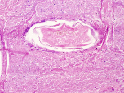
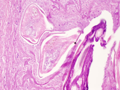
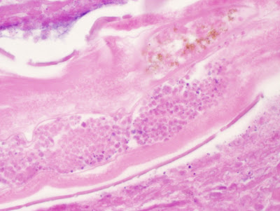
The below photographs are of H&E stained sections from an orchiectomy specimen. The patient is a Californian man with a complaint of "testicular mass." No further history is available. The questions that the referring pathologist has for you are:

1. Is this a worm or some type of artifact?
2. If it is a worm, could it be Strongyloides stercoralis or Toxocara spp.?

What do you think?

(x40 original magnification)


(x100 original magnification)


(x400 original magnification)

(x400 original magnification)

(x400 original magnification)

(x400 original magnification)

(x400 original magnification)


(x1000 original magnification)

(x1000 original magnification)

Sunday, October 16, 2011

Answer to Case 180

Answer: Adult filarial worm, female
Differential diagnosis includes Brugia and Wuchereria species, including zoonotic Brugia.

Additional history was received after the case was reviewed, which included a travel history to Central and South America. Therefore, W. bancrofti is a strong consideration, and serologic testing would be advised. Peripheral blood films could also be identified for microfilariae, although this particular worm does not appear to be gravid and is also somewhat degenerated, indicating that it is no longer viable. For W. bancrofti, blood for microfilariae examination should be obtained between 10 pm and 2 am when the microfilariae are released into the bloodstream.

Anonymous points out "In Pathoparasitology , the classic atlas by Michael Kenney, he says "except for W.bancrofti and aberrant eggs of schistosomes,no parasites are usually found in the testes". Thought this might help."

I agree - this is very helpful for formulating a differential diagnosis. The diagnosis microscopic features include:
1. coiled appearance on cross section with surrounding fibrosis
2. size/diameter
3. suggestion of a double uterus, consistent with an adult female filarial worm

Many times, microfilariae can also be seen within the uterus, although they were not present in this case.

I would like to thanks the excellent DPDx team who helped confirm my initial diagnosis and expand on the differential diagnosis. They have a very useful web site at http://www.dpd.cdc.gov/DPDx/

Monday, October 10, 2011

Case of the Week 179

This week's case is somewhat challenging.

The following objects were seen on a peripheral blood film from a patient who recently immigrated from Africa. They measure 230-240 micrometers in length. No further history is available.
Identification? (CLICK ON IMAGES TO ENLARGE)

original magnification x 400


Original magnification x 1000

Sunday, October 9, 2011

Answer to Case 179

Answer: Loa loa

The key features shown in this case are the presence of a non-staining sheath (below), correct size range, and nuclei that extend to the tip of the tail.


Thanks to everyone who wrote in!

Monday, October 3, 2011

Case of the Week 178

The following is a concentrated stool specimen stained with a modified acid fast stain. The patient had reported to his physician after a week-long bout of diarrhea. He had recently attended an event in which he and other guests had eaten fresh salad. Several of the other guests are also sick.

Objects shown measure 10 micrometers in diameter.
Identification?

Sunday, October 2, 2011

Answer to Case 178

Answer: Cyclospora cayetanesis oocysts

The size, history, and strong acid fast positivity are consistent with a diagnosis of cyclosporiasis. However, one viewer points out that the coccidians are inherently difficult to capture in a single photograph since the cellular features may not be well defined, and that fungal artifacts may still be in the differential. Therefore, I've also included the following wet-prep image showing an oocyst from this same case (CLICK ON IMAGE TO ENLARGE). From this image, the round shape and internal structures can be appreciated.


When examining modified acid fast stains, it is also important to note that the size of the stained objects is also important, since Cryptosporidium spp. oocysts have a very similar appearance, but are smaller (4-6 microns in diameter).

Monday, September 26, 2011

Case of the Week 177

The following images are sections of a formalin-fixed paraffin-embedded gastric biopsy. No further history is available.
(CLICK ON IMAGES TO ENLARGE)

H&E 40 times original magnification


H&E 100 times original magnification


H&E 400 times original magnification

H&E 400 times original magnification

H&E 40 times original magnification


H&E 100 times original magnification


H&E, 400 times original magnification


Trichrome stain, 40 times original magnification


Trichrome stain, 200 times original magnification


Trichrome stain, 400 times original magnification


How would you sign this case out?