A worm-like object was removed during routine colonoscopy from a 60-year old woman and was submitted to surgical pathology for sectioning and staining. Below are representative images from this case. Identification? (CLICK ON IMAGES TO ENLARGE)
H&E, 20x original magnification
H&E, 100x original magnification
H&E, 400x original magnification
H&E, 400x original magnification (with narrowed condenser)
Monday, August 31, 2015
Sunday, August 30, 2015
Answer to Case 362
Answer: Trichuris trichiura (whipworm), cross-sections
There was a lot of great discussion for this case. Multiple readers mentioned the characteristic eggs (described as "lemon", "tea tray" or "football" shaped) with bipolar plugs (arrows below) which are characteristic for T. trichiura. This is best seen by narrowing the condenser - something I teach to all of my students!
Note that some of the eggs resemble those of Enterobius vermicularis, and therefore you would need to consider that parasite in the differential and carefully examine the sections for bipolar plugs.
Arthur Morris also mentions that the low power magnification nicely demonstrates the thin anterior end as well as the thicker posterior end - another diagnostic feature for T. trichiura.
Finally, there is a clearly identifiable stichosome in the anterior portion of the worm - something that I should have highlighted in my initial images - which is also characteristic for T. trichiura and closely related worms (Trichinella, Capillaria).
So as Dan Milner says "this is why we have to crack the 'whip' when teaching pathology! We can't just serve up all this knowledge on a 'tea tray' and expect them to get it. If one can focus on the case and not let the overwhelming pressure of the image 'prolapse' your brain, you'll eventually sort it out."
Or from Florida Fan "Most students need only to learn the simple truth: 'nothing ventured, nothing gained,' and they should love to 'whip' the 'worm' till the foot balls are out, avoiding a 'prolapse at the rear'."
And from Blaine:
This specimen is from the (rectum),
I think it nearly (killed him)
To rock (a worm), that'll make (you squirm)
It's Trichuris trichiura, here we go...
[RUN-DMC fans will get the reference...}
Or for the Star Wars Fans:
Princess Leia was always proud of her Cinnabon hair
Until something similar poked out of her rear
With hyaline bipolar plugs,
This can only be one bug
It's Trichuris trichiura down in the derriere
You are all so creative - I love it.
Sunday, August 23, 2015
Case of the Week 361
The following specimen was submitted to the laboratory for identification (CLICK ON IMAGES TO ENLARGE):
Identification?
How would you go about identification to the species level in your laboratory? (I'm especially interested in hearing what your specific practice is). Thanks!
Using gentle manipulation with forceps, the following were expressed from this object:
Identification?
How would you go about identification to the species level in your laboratory? (I'm especially interested in hearing what your specific practice is). Thanks!
Saturday, August 22, 2015
Answer to Case 361
Answer: Taenia species
Thank you for all of the great responses! Based on the morphology of eggs that were expressed from the tapeworm proglottids, we were able to identify this as a Taenia species. Note that the eggs are round-oval, have a radially-striated wall, and clearly-visible internal hooklets.
As part of this case, I asked readers to comment on which procedure they used in their lab to identify the specific species of Taenia. Several readers commented that they compressed a proglottid between 2 slides (with or without prior clearing with phenol) before trying to count the primary uterine branches off of the central stem. A few mentioned trying to use India ink injection to highlight the branches, although several individuals mentioned how challenging this can be, especially when the proglottids are submitted in formalin. Others mentioned using histopathologic sectioning to visualize the uterine branches.
We've used all of these techniques in my lab, but I have recently settled on histologic sectioning as my method of choice for examining the uterine branches. Even though we have had success with all of the methods mentioned above, I prefer the histologic method for the following reasons:
- It requires the least amount of proglottid manipulation outside of a biosafety cabinet (important since proglottids may contain potentially infectious eggs).
- It doesn't require us to keep phenol or India ink in our lab (two less reagents that require stocking, labelling, monitoring and disposal!)
- Submission in formalin doesn't negatively impact our analysis (and is actually the fixative of choice for histology).
- We have access to an excellent histology lab that can perform the sectioning and staining within 1-2 days (which I feel is acceptable for patient care).
So although we don't play around with the proglottids anymore (which can be fun) we now have a reliable and efficient method for determining the species of Taenia proglottids. Here is an image of the histologic sections from this case (CLICK ON IMAGES TO ENLARGE):
I marked the primary uterine branches in the image below to demonstrate that there are >13 branches present, making this T. saginata/T. asiatica.
Monday, August 10, 2015
Case of the Week 360
This week's case was generously donated by Dr. Sheldon Campbell, "the singing microbiologist." (seriously, his songs are great - check it out here).
This specimen was submitted from a patient in Connecticut.
Identification?
This specimen was submitted from a patient in Connecticut.
Identification?
Sunday, August 9, 2015
Answer to Case 360
Answer: Amblyomma americanum, the Lone Star tick.
An important clue to this case is the presence of a single macula (dot or 'lone star') on the dorsum of the tick from which its name is derived. It is important to keep in mind, however, that other ticks can have maculae on their dorsal surface and therefore characteristics of the the anal groove and mouth parts, as well as the presence or absence of festoons are important for confirming the identification of the tick. Festoons can be particularly challenging to appreciate when the tick is engorged such as in this case, but they can usually be seen using careful examination with a dissecting microscope and lateral light. Below, the presence of faint shadows suggest the presence of festoons (arrow heads).
The CDC has a nice genus-level pictorial tick key that you can access here:
http://www.cdc.gov/nceh/ehs/docs/pictorial_keys/ticks.pdf
Some of you might have been slightly thrown off by the geographic location of this tick (Connecticut). Despite it's name, the Lone Star tick has a distribution far outside of the 'Lone Star state' of Texas. In fact, the range now expands far up into the north east and north central states, and is approaching the upper midwest.
Map from the CDC (http://www.cdc.gov/ticks/maps/lone_star_tick.pdf)
Amblyomma americanum are aggressive biters and are vectors of the organisms causing ehrlichiosis, tularemia and STARI. Their geographic range continues to expand, therefore increasing the number of individuals potentially at risk for A. americanum-transmitted diseases.
Thanks again to Dr. Sheldon Campbell for donating this case.
Monday, August 3, 2015
Case of the Week 359
This week's case is from my archive and therefore it's a little yellow around the edges. However, it's still a neat case that I thought you might like:
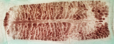
A 3-cm tan-white structure was passed in the stool from a 30 year old woman and submitted for identification. In the lab, the object was cleared and stained using a carmine stain. Identification?
A 3-cm tan-white structure was passed in the stool from a 30 year old woman and submitted for identification. In the lab, the object was cleared and stained using a carmine stain. Identification?
Sunday, August 2, 2015
Answer to Case 359
Answer: Taenia saginata proglottid
As many of you have pointed out, the identification can be made based on the presence of more than 13-15 primary lateral uterine branches on each side of the uterine stem. When counting branches, remember to count them as they come off of the uterine stem rather than at their terminal ends, since the latter will give you a falsely elevated count. Also, remember to only count on 1 side of the uterine stem to get your total. I've highlighted the uterine stem and a few of the primary branches below. Although it's sometimes hard to figure out which are primary vs. secondary branches, I believe we still have at least 13 primary branches in this case (using a conservative estimate).
Of note, Taenia asiatica has a very similar appearance and therefore obtaining a travel history is important in formulating your differential. In my lab, the primary concern is ruling our Taenia solium, since the infected individual and his or her contacts may be at risk for cysticercosis from eggs being shed in the stool. In comparison, the eggs of T. saginata and T. asiatica are not infectious to humans and do not cause cysticercosis.
Subscribe to:
Posts (Atom)


