Sunday, May 18, 2025

Case of the Week 777

 Dear Readers,

I am excited to announce that we are celebrating our 777th case! 

In honor of this milestone, we have a selection of 3 helminth eggs for you to identify. You win the parasite jackpot if you can get all three. There is an 'easy' and 'hard' version, so you get to take your pick.

EASY VERSION

Egg 1 (50 micrometers long)
Egg 2 (55 micrometers long)
Egg 3 (35 micrometers long)

HARD VERSION

Egg 1 (45 micrometers in diameter)


Egg 2 (95 micrometers long)

Egg 3 (One of 15 objects freed from a 4mm structure)


Saturday, May 17, 2025

Answer to Case 776

 Answer to the Parasite Case of the Week 776: Toxoplasma gondii tachyzoites. As the name implies, the tachyzoite is the rapidly dividing stage of the parasite (tachy is from the Greek takhus meaning rapid, swift). Tachyzoites invade cells and divide rapidly within parasitophorous vacuoles, ultimately rupturing infected cells. Tachyzoites divide by endodyogeny, an interesting form of replication seen with some coccidia in which two daughter cells develop internally within the parent cell without nuclear conjugation. The parent cell is consumed in the process - yikes!

Shown here is the classic arc-shaped tachyzoite and 2 daughter cells resulting from the process of endodyogeny:


Monday, May 12, 2025

Case of the Week 776

This week's case is a brain biopsy from a middle-aged man with untreated HIV. The specimen appeared necrotic and bloody. Touch preps were made from the material and stained with Giemsa. From the images below taken with the 100x oil objective, what is your diagnosis?


Tuesday, May 6, 2025

Case of the Week 775

This week's case was inspired by Dr. Eric Rosenbaum. Several objects were submitted for parasite identification, including tan-white hair-like structures measuring 30 mm long by <0.2 mm wide. Here is their appearance by light microscopy. How would you sign this case out?


Sunday, May 4, 2025

Answer to Case 775

Answer to the Parasite Case of the Week 775: Not a parasite. This is actually a hair from my house cat, Walter. Here he is keeping me company while I am trying to update a parasitology chapter:

As noted by Florida Fan, "The objects have neither external nor internal organs. We have a common saying: "No head, no tail, no guts, no parasites”

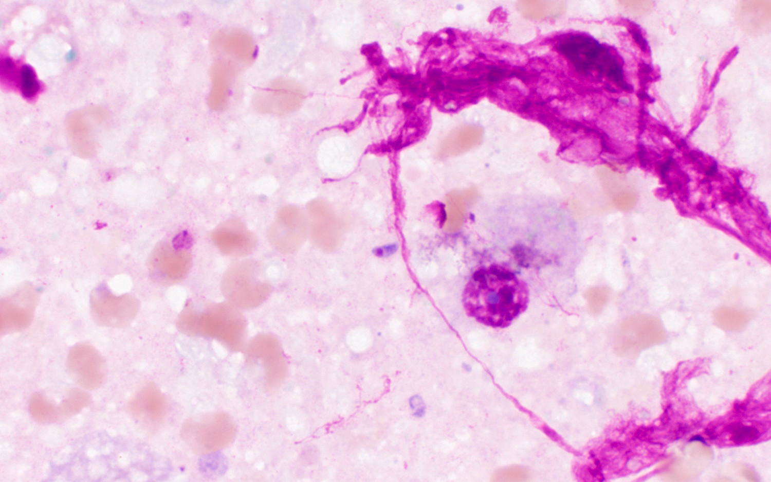
Most readers realized that this object was not a parasite, but a few did suggest that it was a tapeworm such as Rodentolepis (formerly Hymenolepis) nana, given the superficial resemblance of the cuticle scale and fragmented, 'ladder like' medulla to craspedote (overlapping) proglottids. However, we can differentiate the hair from R. nana proglottids by the smaller size, refractile nature, and lack of uterine structures. Check out the following comparison of our current case to an image of R. nana from a previous post donated by Dr. Emily Snaverly:
A few particularly savvy readers correctly guessed the nature of this object. There are a number of animal hairs that have this appearance, including some rodents (e.g., mice, chinchillas). You can see an amazing archive of mammalian hair images HERE. From the standpoint of the diagnostic laboratory, the important thing is to recognize that the submitted object is not a parasite and report it as such.