Answer: Ascaris lumbricoides and Trichuris trichiura eggs.
As noted by a few of the readers, both fertilized and unfertilized Ascaris ova are present. It is common to see co-infections with geohelminths in patients from endemic settings. Therefore, a good take home point is: if you see one parasite, keep looking!
Sunday, July 13, 2014


Monday, July 7, 2014
Case of the week 310
I have an unusual case for you this week. The following images are of a stool agar culture plate for Strongyloides stercoralis after 6 days of incubation at room temperature:


Using a standard light microscope, the following video was captured using the 10x objective (100x total magnification):
Note that there is subtle movement in these filamentous structures (the choppiness is an artifact of my video camera).
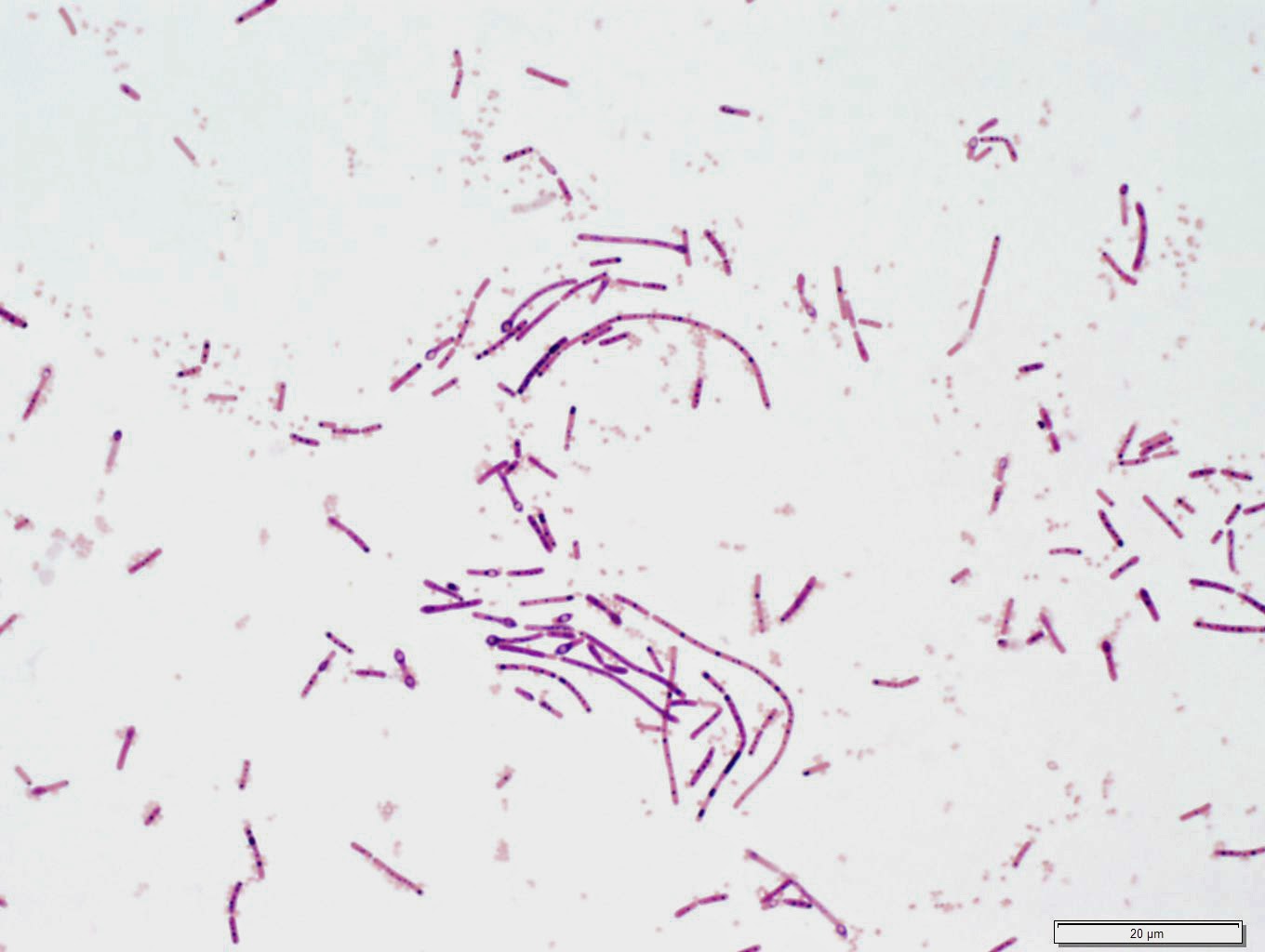
To further investigate this, we performed a Gram stain of material taken from the fringed edge:
Gram at 100x

Gram at 1000x
Identification? Suggested further work-up?
Using a standard light microscope, the following video was captured using the 10x objective (100x total magnification):
Note that there is subtle movement in these filamentous structures (the choppiness is an artifact of my video camera).
To further investigate this, we performed a Gram stain of material taken from the fringed edge:
Gram at 100x
Gram at 1000x

Identification? Suggested further work-up?
Sunday, July 6, 2014
Answer to Case 310
Answer: No Strongyloides stercoralis or other larvae detected. A motile Bacillus sp. was present, accounting for the motility observed on the Strongyloides agar culture plate.
This was an unusual case because the bacteria produced an interesting motion which superficially resembled larval movement. However, the observed motility was not the expected sinuous movement of larvae through bacteria, but instead, consisted of regular and continuous clockwise or counterclockwise swirling within a confined region of the plate.
Multiple Gram stained preparations from these areas showed large Gram-variable to Gram positive rods with spores consistent with a Bacillus species. Smaller Gram negative bacilli (likely coliforms) were also seen in the background.
It's important to remember that things other than Strongyloides stercoralis larvae can demonstrate motility on Strongyloides agar cultures. In this case, the unusual motility pattern was due to fecal bacteria. However, we've also seen mites, hookworm larvae, and even larvae from free-living nematodes demonstrate visible motility. That is why we work up all positive cultures with microscopic identification of the motile objects.
Thanks for the great discussion on this one!
This was an unusual case because the bacteria produced an interesting motion which superficially resembled larval movement. However, the observed motility was not the expected sinuous movement of larvae through bacteria, but instead, consisted of regular and continuous clockwise or counterclockwise swirling within a confined region of the plate.
Multiple Gram stained preparations from these areas showed large Gram-variable to Gram positive rods with spores consistent with a Bacillus species. Smaller Gram negative bacilli (likely coliforms) were also seen in the background.
It's important to remember that things other than Strongyloides stercoralis larvae can demonstrate motility on Strongyloides agar cultures. In this case, the unusual motility pattern was due to fecal bacteria. However, we've also seen mites, hookworm larvae, and even larvae from free-living nematodes demonstrate visible motility. That is why we work up all positive cultures with microscopic identification of the motile objects.
Thanks for the great discussion on this one!
Monday, June 30, 2014
Case of the Week 309
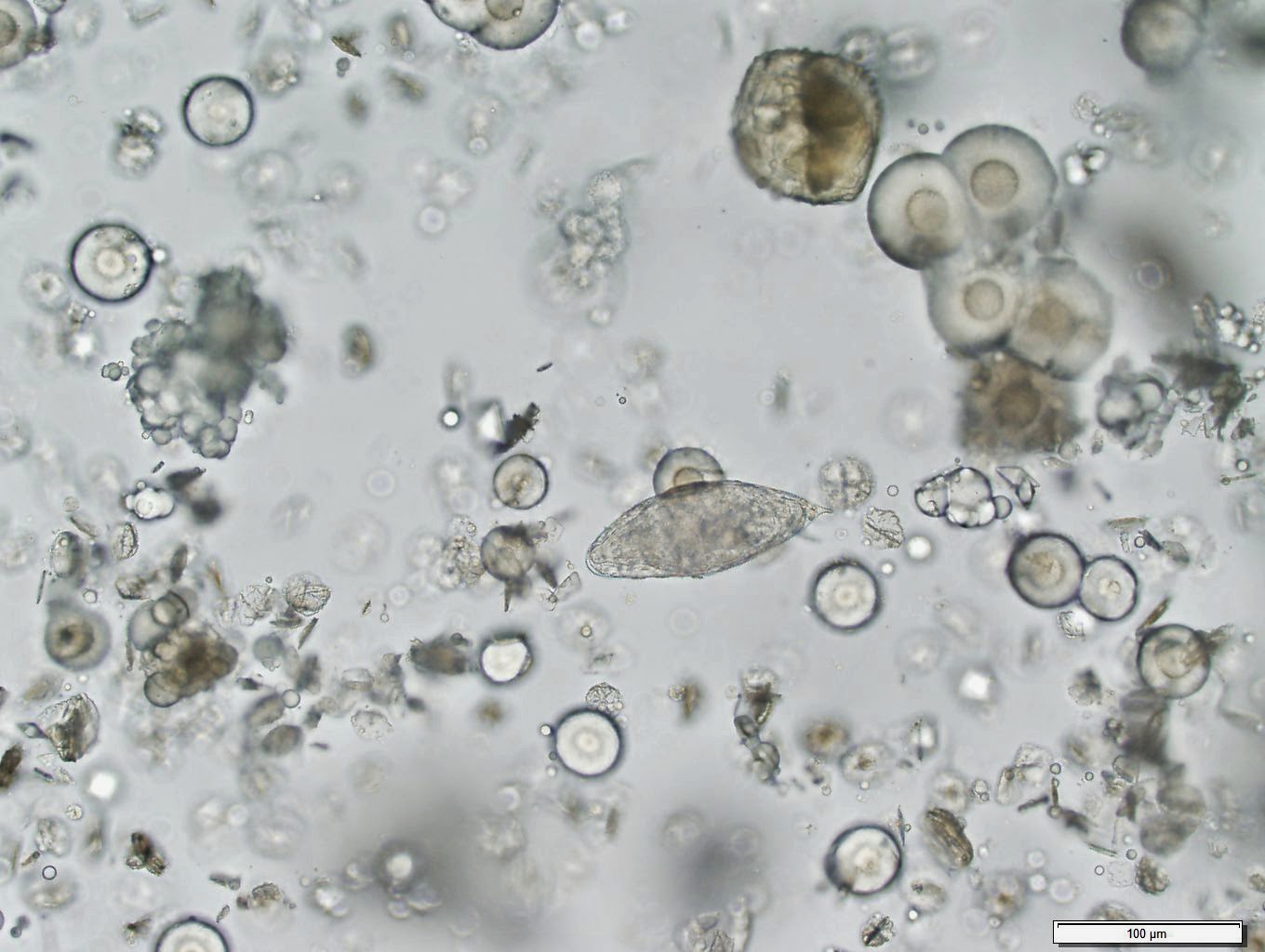
The following structures were seen in a refrigerated urine specimen. Identification?


Sunday, June 29, 2014
Answer to Case 309
Answer: Schistosoma haematobium ova/eggs
Everyone who wrote in had the correct answer. These eggs have a very characteristic appearance with a large size (112 - 170 micrometers in length) and terminal spine. As mentioned by Hans, you can check to see if the eggs are still viable by looking for the internal beating flame cells, particularly in fresh specimens. Unfortunately this specimen was several days old by the time it had reached us (it had been sent in from another lab as a consult) and no motility was observed - possibly due to the age of the specimen. Hans also mentions that you can hatch the eggs - that's always something fun to do!
Anon noted the considerable debris that was present in the background. Indeed, one of the reasons I chose this case for a blog post is because it had a lot of crystals in the background which can make it challenging to spot the eggs. This is seen occasionally with refrigerated specimens; gently heating them will often dissolve the crystals and allow the urine to be examined for parasite eggs. Some types of urine crystals are commonly seen and do not necessarily indicate disease. However, other types of crystals are strongly associated with disease - particularly when persistent. When in doubt, consult your Clinical Chemist or suggest that a urine specimen be sent for crystal analysis.

Everyone who wrote in had the correct answer. These eggs have a very characteristic appearance with a large size (112 - 170 micrometers in length) and terminal spine. As mentioned by Hans, you can check to see if the eggs are still viable by looking for the internal beating flame cells, particularly in fresh specimens. Unfortunately this specimen was several days old by the time it had reached us (it had been sent in from another lab as a consult) and no motility was observed - possibly due to the age of the specimen. Hans also mentions that you can hatch the eggs - that's always something fun to do!
Anon noted the considerable debris that was present in the background. Indeed, one of the reasons I chose this case for a blog post is because it had a lot of crystals in the background which can make it challenging to spot the eggs. This is seen occasionally with refrigerated specimens; gently heating them will often dissolve the crystals and allow the urine to be examined for parasite eggs. Some types of urine crystals are commonly seen and do not necessarily indicate disease. However, other types of crystals are strongly associated with disease - particularly when persistent. When in doubt, consult your Clinical Chemist or suggest that a urine specimen be sent for crystal analysis.
Sunday, June 22, 2014
Case of the Week 308
The following case was generously donated by FloridaFan.
The patient is an 85 year old woman in a skilled nursing facility. She experienced a rash involving her scalp, and so scrapings were obtained and sent to the laboratory. Identification?


The patient is an 85 year old woman in a skilled nursing facility. She experienced a rash involving her scalp, and so scrapings were obtained and sent to the laboratory. Identification?
Saturday, June 21, 2014
Answer to Case 308
Answer: Demodex sp. and Sarcoptes scabei
This was a fun case because of the presence of 2 different mites in a single specimen. As noted by Arthur Morris, "it's likely that Sarcoptes is causing the rash, though in some cases Demodex can cause Sarcoptes-like rashes."
Mites belong to the class Arachnida, and therefore have 8 legs in the nymph and adult forms, but only 6 legs in the larval form, when they are newly emerged from the egg. Although it's a bit hard to make out in the photograph, I believe we have a larval Sarcoptes scabei mite in the bottom left of the image below, which helps explain its small size.

Thanks again to FloridaFan for donating this great case.
This was a fun case because of the presence of 2 different mites in a single specimen. As noted by Arthur Morris, "it's likely that Sarcoptes is causing the rash, though in some cases Demodex can cause Sarcoptes-like rashes."
Mites belong to the class Arachnida, and therefore have 8 legs in the nymph and adult forms, but only 6 legs in the larval form, when they are newly emerged from the egg. Although it's a bit hard to make out in the photograph, I believe we have a larval Sarcoptes scabei mite in the bottom left of the image below, which helps explain its small size.
Thanks again to FloridaFan for donating this great case.
Thursday, June 12, 2014
Case of the Week 307
The following were removed from a wound and submitted to the laboratory for identification.
(ruler marks below represent millimeters)


Many were still moving!
The following is from the posterior section.

Identification?
(ruler marks below represent millimeters)
Many were still moving!
The following is from the posterior section.
Identification?
Wednesday, June 11, 2014
Answer to Case 307
Answer: Fly larvae (maggots). You can tell that these are second instars because they only have 2 spiracular slits rather than 3. It is difficult to identify early instars to the genus level, given the lack of available literature. For the 3rd instar larvae, important identifying features include the mouth parts, posterior spiracles, body shape and arrangement of cuticular spines. You can often view the spiracles using only a stereomicroscope, although in this case, I cut off the posterior end and mounted it on a glass slide.

In this case, I can tell you that these are specifically blowfly larvae; Lucilia (Phaenicia) sericata; also sold as Medical Maggots (TM), since they were purchased by my institution. These maggots are actually cleared by the FDA for "debriding chronic wounds such as pressure ulcers, venous stasis ulcers, neuropathic foot ulcers and non-healing traumatic or post surgical wounds." Since they ingest only dead tissue, they offer a very effective means of wound debridement.
When ordered, they come in a small gauze, which is applied directly to the wound and then covered with a dressing. The instructions warn that after consuming the dead tissue, the maggots may migrate outside of the dressing, which can be a significant cause of consternation for the patient and nursing staff! Here is a link to some great information on maggot therapy.
In this case, I can tell you that these are specifically blowfly larvae; Lucilia (Phaenicia) sericata; also sold as Medical Maggots (TM), since they were purchased by my institution. These maggots are actually cleared by the FDA for "debriding chronic wounds such as pressure ulcers, venous stasis ulcers, neuropathic foot ulcers and non-healing traumatic or post surgical wounds." Since they ingest only dead tissue, they offer a very effective means of wound debridement.
When ordered, they come in a small gauze, which is applied directly to the wound and then covered with a dressing. The instructions warn that after consuming the dead tissue, the maggots may migrate outside of the dressing, which can be a significant cause of consternation for the patient and nursing staff! Here is a link to some great information on maggot therapy.
Sunday, June 1, 2014
Case of the Week 306
The following case was generously donated by Dr. Tom Davis and Dr. Ryan Relich (a.k.a. MicrobeMan).
This was an incidental finding during colonoscopy and polypectomy in a 60-year old female of unknown nationality and travel history. Identification?
Hematoxylin and Eosin


This was an incidental finding during colonoscopy and polypectomy in a 60-year old female of unknown nationality and travel history. Identification?
Hematoxylin and Eosin
Subscribe to:
Posts (Atom)


