Answer: Larval ticks, Ixodes species
These little guys were a bit hard to make out due to their small size, but you can nicely see several characteristic features, including 6 legs (versus the 8 legs of nymphs and adults), and - if you squint - a U-shaped anal groove, consistent with Ixodes species.
As mentioned by FP in Burlington, VT, Borrelia burgdorferi, the agent of Lyme disease, is not transmitted tranovarially from mother to larva. Therefore, larvae are not infected and cannot transmit this bacterium to humans. Larvae can become infected when they take their first blood meal, after which they molt into nymphs. Therefore, unlike larvae, nymphs ARE capable of transmitting B. burgdorferi to humans, and present a particular hazard due to their small size which makes them easy to overlook.
Saturday, August 23, 2014
Monday, August 18, 2014
Case of the Week 315
Sunday, August 17, 2014
Answer to Case 315
Answer: Moniliformis moniliformis, one of the principle agents of acanthocephaliasis.
The adult worm has a very characteristic morphologic appearance, which Heather A. aptly describes as resembling a "bendy straw" (see Here for information about bendy straws). Not shown here (since it was retracted into the worm) is the hooked proboscis from which the acanthocephalans get their name (Acanth is Greek for spiny or thorny).
The eggs are also characteristic, with a relatively large size (90-120 micrometers long) and internal larva with rostellar hooks.
Humans are accidental hosts, usually acquiring infection via ingestion of an infected insect such as a cockroach or beetle. Moniliformis moniliformis does not always mature in humans, and when it does, it seldom produces eggs. Therefore, this case is interesting in that a mature and gravid female was identified.
The adult worm has a very characteristic morphologic appearance, which Heather A. aptly describes as resembling a "bendy straw" (see Here for information about bendy straws). Not shown here (since it was retracted into the worm) is the hooked proboscis from which the acanthocephalans get their name (Acanth is Greek for spiny or thorny).
The eggs are also characteristic, with a relatively large size (90-120 micrometers long) and internal larva with rostellar hooks.
Humans are accidental hosts, usually acquiring infection via ingestion of an infected insect such as a cockroach or beetle. Moniliformis moniliformis does not always mature in humans, and when it does, it seldom produces eggs. Therefore, this case is interesting in that a mature and gravid female was identified.
Monday, August 11, 2014
Case of the Week 314
Sunday, August 10, 2014
Answer to Case 314
Answer: Not a parasite: uric acid crystals
Crystals are commonly seen in urine specimens and can be a very good mimic for parasite eggs. This is particularly true for the 'lemon-shaped' uric acid crystals which mimic Schistosoma haematobium eggs. You can differentiate uric acid crystals from S. haematobium eggs by the following features:
1. Uric acid crystals vary in size and shape and are often much smaller than S. haematobium eggs. In contrast, S. haematobium eggs are regular in size and shape, and quite large (approximately 150 micrometers in length).
2. Uric acid crystals commonly have points on both ends instead of the single 'pinched-off' spine of S. haematobium eggs. They can also have lateral points or take on other shapes.
3. There are no internal parasite structures in crystals
4. Finally, crystals often fracture and break, although this is not shown in this case.
Here is a picture from this case that demonstrates many of these features, including points at both ends, variability in size and shape and lack of internal parasite structures.
Thank you all for writing in on this case! Crystals can be challenging, so I like to show them whenever they come up in my lab.
Crystals are commonly seen in urine specimens and can be a very good mimic for parasite eggs. This is particularly true for the 'lemon-shaped' uric acid crystals which mimic Schistosoma haematobium eggs. You can differentiate uric acid crystals from S. haematobium eggs by the following features:
1. Uric acid crystals vary in size and shape and are often much smaller than S. haematobium eggs. In contrast, S. haematobium eggs are regular in size and shape, and quite large (approximately 150 micrometers in length).
2. Uric acid crystals commonly have points on both ends instead of the single 'pinched-off' spine of S. haematobium eggs. They can also have lateral points or take on other shapes.
3. There are no internal parasite structures in crystals
4. Finally, crystals often fracture and break, although this is not shown in this case.
Here is a picture from this case that demonstrates many of these features, including points at both ends, variability in size and shape and lack of internal parasite structures.
Thank you all for writing in on this case! Crystals can be challenging, so I like to show them whenever they come up in my lab.
Monday, August 4, 2014
Case of the Week 313
The following case was generously donated by Florida Fan. These images were all obtained from specimens submitted from a 5-year old girl who was possibly the daughter of a migrant farmer. Identification? Bonus: name the forms!
Sunday, August 3, 2014
Answer to Case 313
Answer: Pediculus humanus capitis
All that wrote in got this correct: This case donated by Florida Fan nicely demonstrates 4 different stages/sexes; in order of presentation:
Adult male
Adult female
Eggs (nit) cemented onto a hair shaft
and last but not least, a nymph hatching from an egg!
All that wrote in got this correct: This case donated by Florida Fan nicely demonstrates 4 different stages/sexes; in order of presentation:
Adult male
Adult female
Eggs (nit) cemented onto a hair shaft
and last but not least, a nymph hatching from an egg!
Monday, July 21, 2014
Case of the Week 312
Dear Readers,
I'd like to first thank This Week in Parasitism and the hosts, Vincent Racaniello and Dickson Despommier for featuring my blog as their 75th podcast. You can hear the podcast HERE. Please check out their other podcasts - they're fun and contain great information!
Now for our case this week: A peripheral blood thin film from a patient with recurring fevers and recent travel to India. Identification?

I'd like to first thank This Week in Parasitism and the hosts, Vincent Racaniello and Dickson Despommier for featuring my blog as their 75th podcast. You can hear the podcast HERE. Please check out their other podcasts - they're fun and contain great information!
Now for our case this week: A peripheral blood thin film from a patient with recurring fevers and recent travel to India. Identification?

Sunday, July 20, 2014
Answer to Case 312
Answer: Plasmodium vivax
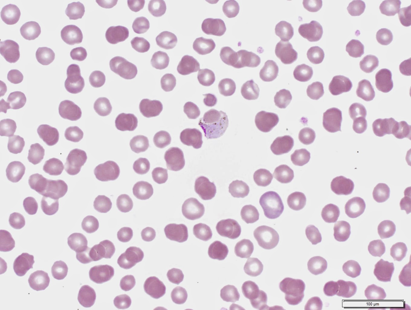
As described by Florida Fan, many of the characteristic P. vivax morphologic features are present in these images including infected RBCs that are larger than non-infected RBCs, schizonts with many merozoites (12-24) and infected cell "hugging" of adjacent RBCs. In fact, this case had the "huggiest" (is that a word?) RBC I've ever seen. This case also had free merozoites which are not commonly seen in peripheral blood smears. Lastly, faint stippling (Schuffner's dots) was seen in some cells. Here are some representative images from this case:
Monday, July 14, 2014
Case of the Week 311
Here's a nice straight-forward case this week. The following were seen on Ova and Parasite exam performed on an adopted child from Ethiopia. Identification? (CLICK ON THE IMAGES TO ENLARGE)
Subscribe to:
Posts (Atom)