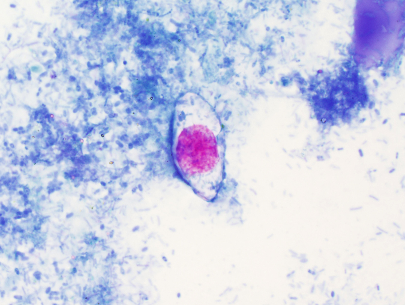
This week's case is from my own lab, with the beautiful images taken by Emily F. The patient is an asymptomatic male who underwent routine screening colonoscopy, which revealed the following:
Two objects were removed during colonoscopy and submitted to the Clinical Parasitology lab for identification:
By manipulating the objects, we were able to express the following:
Identification?
Monday, July 18, 2016
Sunday, July 17, 2016
Answer to Case 405
Answer: Trichuris trichiura (whipworm)
As pointed out by Arthur V and the other readers, the shape of the adult worms (narrow anterior end, broad posterior end) and the eggs are characteristic for this roundworm. Note that there is both a female (second image, straight posterior) and a male (image, curved posterior) in this specimen.
Another interesting feature of this case is that we were able to express both immature and mature eggs out of the female worm using blunt manipulation. The differences in maturity are responsible for the slightly different appearances of the eggs (kudos to the readers that pointed this out!) Here are the 2 eggs side-by-side for comparison:
Sunday, July 10, 2016
Case of the Week 404
This week's case was generously donated by Florida Fan. This structure was found in a concentrated stool specimen from a teenage male. It measures approximately 45 micrometers in diameter (shown at 400x total magnification). Identification?
Saturday, July 9, 2016
Answer to Case 404
Answer: Hymenolepis nana egg
As pointed out by Sheldon, BW, and Anon, this is a beautiful example of an H. nana egg, with refractile internal hooklets and 2 clearly visible polar thickenings. The arrows on the image below point out the polar thickening from which the polar filaments arise.

Thanks again for Florida Fan for donating this great case!
Sunday, July 3, 2016
Case of the Week 403
Happy Independence day to all of my American readers! Here is some red (magenta), white and blue for you - found in a modified acid fast-stained stool specimen:
The objects measure approximately 35 micrometers in length. Identification?
Saturday, July 2, 2016
Answer to Case 403
Answer: Cystoisospora (formerly Isospora) belli
As pointed out by the readers, these are immature oocysts, since only 1 sporoblast is visible.
This is the state that this organism is shed in the stool of infected individuals. The oocysts then mature in the environment into their infective form, with the sporoblast dividing into 2 sporoblasts. The sporoblasts develop cell walls and become sporocysts containing 4 infective sporozoites each.
Thanks to Florida Fan for pointing out that the modified/hot safranin stain will also beautifully stain the oocysts of Cystoisospora (and other coccidia). The oocysts also exhibit striking autofluorescence using UV microscopy (excitation filter at 330-365 nm or 450-490 nm).
I hope you all had a safe and happy July 4th weekend!
Monday, June 27, 2016
Case of the Week 402
This week's case was generously donated by Dr. Rocco LaSala. The specimen was retrieved from the stool of a 70 year-old-woman with approximately 12 months of diarrhea, abdominal cramping, and unintended weight loss (> 40 lbs). The structures are thin, brown, and 'thread-like', measuring approximately 0.7 mm in width.
Specimens were also submitted for histology, which showed the following on H&E staining:
Identification?
Sunday, June 26, 2016
Answer to Case 402
Answer: Not a parasite; plant material.
This case was clearly not a parasite (or even animal tissue), as seen on both gross and microscopic images. The material most closely resembles banana seeds, as seen on a previous case that I've posted.
Unfortunately, this doesn't help elucidate the cause of the patient's symptoms, and therefore additional studies will be necessary.
Thanks again to Rocco for sharing this case with us.
Tuesday, June 21, 2016
Case of the Week 401
This week's case is in honor of Father's Day, which was just 2 days ago, as well as Mother's Day, which was in May.
The following were submitted to my laboratory for identification. The larger of the 2 arthropods was still alive. Identification?
Thanks to Emily Fernholz for taking these gorgeous photos!
The following were submitted to my laboratory for identification. The larger of the 2 arthropods was still alive. Identification?
Here are some close-up shots:
Thanks to Emily Fernholz for taking these gorgeous photos!
Monday, June 20, 2016
Answer to Case 401
Answer: Dermacentor sp. ticks: an adult male and female (latter is heavily engorged) and newly-laid eggs.
We were very excited to find these eggs in the container along with the live female. Therefore, we decided to try to hatch them. Special thanks to William Nicholson and Graham Hickling who provided the necessary instructions, and to Jim, Lynn, Heather, Emily and the rest of my awesome lab staff who made it all happen. It turns out that hatching larvae from eggs is not a straight-forward process. The eggs need to be maintained around room temperature, and importantly, need to be kept in a humid environment (e.g. 80-90%). Also, they need to be monitored regularly for fungal growth which could kill them (I can provide instructions to anyone who is interested). To accomplish this, we set up the following humidity chamber:
We went through a couple of different iterations of the humidity chamber, using different solutions and containers, but eventually settled on the design shown here - a simple sealed Tupperware container with distilled water at the base. The tick eggs are in a Petri dish sealed with gas-permeable tape and elevated above the water on a used pipette tip holder).
We then patiently waited for several weeks, monitoring the eggs closely.
As a reminder, here is what the eggs shortly after being laid (May 20):
We were excited to see that they appeared to mature after ~4.5 weeks of incubation (June 17):
By June 23rd, they looked ready to hatch:
And sure enough, by the next day we had hatching larvae!

So now you may be wondering what we are going to do with all of them? Well, as cute as they are, they're still baby ticks that could be harboring human pathogens, so they will be dumped into formalin to be preserved for posterity.
Subscribe to:
Posts (Atom)