Wednesday, May 18, 2022

Case of the Week 682

This week we have a special guest author, Hadel Go, a medical student (Class of 2024) who is greatly interested in parasitology. I'm very excited that Hadel will be our first ever guest author! The following is our case that she will be teaching us about later this week.

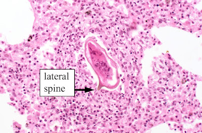
The patient is a young man from Madagascar who presented with a 1-week history of chest pain, night sweats, and fever. Chest X-ray showed a right upper lobe peripheral cavitary lesion. Work up for tuberculosis including AFB stained smears of sputum and sputum PCR using the Xpert MTB/RIF assay were negative. Therefore, a wedge resection of the abscess was performed and revealed the following (H&E stain, 400x original magnification):

What is your diagnosis?


Tuesday, May 17, 2022

Answer to Case 682

 Answer to the Parasite Case of the Week 682: Schistosoma mansoni egg.

The following post is from our first ever guest author, Hadel Go. I think you will all agree that Hadel did an outstanding job writing up the answer to the case of the week, and that this is truly one of the best case answers we have had on this blog.

__________________________________

Hadel Go, Medical Student, Guest Author

This is a Schistosoma mansoni egg in lung tissue as many of you correctly identified in the comments. The large lateral spine is a dead giveaway and creates the “quote bubble” morphology mentioned by Jacob @eternalstudying on Twitter. See more images here: Schistosoma mansoni, eggs, tissue (parasitewonders.com).
Here is an image of a S. mansoni egg in an unstained stool wet preparation.
The burning question in this case is: what is a Schistosoma mansoni egg doing in the lung? Typically, S. mansoni eggs are found in the stool, intestinal tract, and liver of infected humans. Paired adults (male and female) anchor themselves in a venule of the intestinal wall (often in the inferior mesenteric veins) and release eggs. Some of these eventually make their way through the intestinal wall into the lumen and out of the body with the stool, thus allowing the parasite's life cycle to continue. However, many of the eggs remain in the venule and travel via the portal circulation to the liver, where they become lodged and cause granulomatous inflammation.

Chronic infections and high parasite burden can induce portal hypertension due to obstruction by granulomas. This results in shunting and embolization of the eggs in ectopic sites outside the liver and intestines. 

Eggs can also occasionally be seen in the lungs during the acute stage of infection, during which larval migration, maturation and initial egg production occurs. Symptoms of acute pulmonary schistosomiasis occur within 2-8 weeks after exposure and patients present with a dry cough, dyspnea, wheeze, night sweats, or chest pain (as seen in this patient). Chest x-ray and CT imaging of the lungs will usually show small bilateral ground glass nodules measuring 2-15mm. Complications can include pulmonary arteritis and cor pulmonale. 

DID YOU KNOW?
Schistosomiasis is also known as bilharzia, named after the German physician and parasitologist Theodor Bilharz. It is a systemic hypersensitivity reaction to the parasite eggs that occur weeks after the initial infection. Acute symptoms include fever (Katayama fever), cough, abdominal pain, diarrhea, hepatosplenomegaly and eosinophilia. Madagascar has the fifth highest rate of schistosomiasis in the world.

LIFE CYCLE
1. Eggs in stool from infected humans are shed in water and hatch miracidia
2. Miracidia enter Biomphalaria snail tissue and develop sporocysts
3. Free swimming cercariae from snails are released in water
4. Cercariae penetrate human skin, lose their tails, and become schistosomulae
5. Schistosomulae spend a few days in the skin before entering the lymphatics and venules
6. On day 5-7, they migrate to the lungs and into the left side of the heart
7. After >15 days, they reach the portal blood in the liver
8. In the hepatoportal circulation, they meet their partner (they’re monogamous!)
9. They mature into adults and the pair migrate to the inferior mesenteric venules and lay eggs

FUN FACTS
Egg Size: 115–175 µm long and 45–47 µm wide, 150 µm diameter on average
Adult Male: 0.6-1.1cm long, 0.1cm wide; white
Adult Female: 1.2-1.6cm long, 0.016 wide (thinner and longer); darker in color due to hemozoin pigment
Distribution: sub-Saharan Africa, Brazil, Venezuela, Suriname, Caribbean, Arabian peninsula
Intermediate host: Biomphalaria snails
Treatment: Praziquantel (reinfection after treatment is common)

TAXONOMY
Kingdom: Animalia
Phylum: Platyhelminthes
Order: Diplostomida
Family: Schistosomatidae

More information:
1. CDC - DPDx - Schistosomiasis Infection
2. Pulmonary Schistosomiasis – Imaging Features - PMC (nih.gov)
3. Schistosoma mansoni Eggs in Spleen and Lungs, Mimicking Other Diseases - PMC (nih.gov)
4. Frontiers | Schistosomes in the Lung: Immunobiology and Opportunity | Immunology (frontiersin.org)
5. How IVI is confronting schistosomiasis in Madagascar - IVI
6. Madagascar.pdf (stanford.edu)
7. Schistosomiasis (1990): Entire title - YouTube


Tuesday, May 10, 2022

Case of the Week 681

This week's case was generously donated by Tracie Rose from Seattle. The following objects were seen in a Papanicolaou-stained preparation of aspirate fluid from a liver cyst. the patient is from Afghanistan. 


Identification? 

Monday, May 9, 2022

Answer to Case 681

 Answer: Echinococcus sp. invaginated protoscolices

Given the clinical history of a cystic lesion in a patient from Afghanistan, the causative agent is probably E. granulosus. 

The protoscolices are somewhat degenerated, but you can still make out the internal row of hooklets:

The protoscolex will evert if ingested by the canine definitive host and form the scolex of the adult tapeworm. Thanks again to Tracie Rose and her lab for donating this case!

If you liked seeing parasites stained with the Papanicolaou (pap) stain, you can also check out this previous case of Schistosoma haematobium eggs in urine. The pap stain is really quite beautiful!

Monday, May 2, 2022

Case of the Week 680

 This week's case is from Drs. Harsha Sheorey and Lauren McShane from Australia. The following were seen in bronchial washings from an immunocompromised patient (fluorescent prep for fungi and wet preps):


Here are the culture plates from the bronchial washings.


The patient also has Gram negative bacteremia. 

Diagnosis? As a bonus, what forms are we seeing in the respiratory specimens?


Sunday, May 1, 2022

Answer to Case 680

 Answer: Strongyloides stercoralis hyperinfection

This impressive case shows numerous L3 (or possibly L4) S. stercoralis larvae from bronchial washings, and the accompanying culture plates showing bacterial colonies growing in the wake of migrating larvae. 


Thanks again to Harsha and Lauren for donating this case!

Wednesday, April 13, 2022

Case of the Week 679

Here is a beautiful tick for you all to identify from Florida Fan - something that is very timely for this season! You can use this TICK KEY to help you get it into the correct genus. What is the potential significance of this finding?


Tuesday, April 12, 2022

Answer to Case 679

 Answer to Parasite Case of the Week 679: Haemaphysalis sp. 

Congratulations to Blaine Mathison, Khaled Itani, Olugbenga Samuel Babatunde, Souti Prasad Sarkhel, Robyn Nadolny, Marc Couturier, @TickReport, and Alvaro Faccini-Martinez for the correct identification! 

It can be tricky to differentiate Haemaphysalis from from Rhipicephalus, so I created the following pictorial guide to help illustrate the key features. 


In the end, it all comes down to a difference of angles - in Haemaphysalis, the outward facing angle is from the palps, whereas with Rhipicephalus, it's from the basis capituli. Both of these ticks also have festoons and their mouthparts are as long as the basis capituli, so they otherwise have a very similar appearance. 

As Blaine points out, there are several members of this genus that are found in the United States, including the newly-invasive H. longicornis, or Asian longhorned tick. There arent' enough features shown in this case to get to the species level, but if you ever have a specimen in your laboratory, you can use this recently published KEY for assistance. Also, if you'd like to learn more about H. longicornis, the CDC has a brief overview HERE. The USDA also has a great site on H. longicornis that includes a regularly-updated map of the tick's distribution throughout the United States.

Thanks again to Florida Fan for donating this case!

Tuesday, April 5, 2022

Case of the Week 678

This week's fascinating and unusual case was donated by Dr. Vicki Schnadig at University of Texas Medical Branch. The patient is an elderly woman from rural Louisiana who presented with a subcutaneous thigh nodule. Dr. Schnadig's team first performed a fine needle aspirate of the lesion which showed the following laminated spherical structure, but no definitive pathogen:

Following this, the team noted a worm-like object protruding from the aspiration site and pulled it out:

The following are histologic sections of this object:


Diagnosis?


Sunday, April 3, 2022

Answer to Case 678

 Answer to the Parasite Case of the Week 678: Sparganosis - infection with the larval form (sparganum) of Spirometra sp.  Kudos to Dr. Francisco Bravo for getting the correct answer, and for the nice description from Florida Fan.

This was a fascinating case as sparganosis is a relatively rare parasitic infection in humans, and the way it was discovered following fine needle aspiration is very unique. It's possible that the laminated calcification seen on the aspirate smear is a calcareous corpuscle, but it's hard to know for sure. Therefore, it was a good thing that the sparganum began to extrude from the aspirate site and could be removed. It's macroscopic and microscopic appearance are diagnostic. Note the longitudinal smooth muscle fibers, loose stroma, and numerous calcareous corpuscles. The latter are seen in all cestodes, in both larval and adult forms. In this case, they mostly appear as empty oval-shaped structures, but can also calcify and appear dark purple on routine H&E stain.

Although removal is curative, this patient should be counseled to avoid eating undercooked fish, reptiles and frogs, to avoid acquiring another case of sparganosis in the future!
