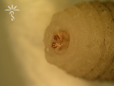
This week's case is by Idzi Potters and the Institute of Tropical Medicine, Antwerp. The following object was removed from a furuncular skin lesion in a patient with recent travel to Uganda. Once they were able to keep it still (!), Idzi captured the following photographs demonstrating all of the diagnostic features. What is your identification?
Thursday, December 7, 2023
Sunday, December 3, 2023
Answer to Case 733
Answer to the Parasite Case of the Week 733: Cordylobia anthropophaga, myiasis causing fly larva.
Florida Fan painted an accurate picture of this parasite with his vivid description: "The mango fly, aka Tumbu fly, lays eggs on human clothing hung to dry in the sun. The Man-Eating larvae hatching eats its way into the skin of its prey. The three sinuous slits are definite identification clues. Ironing the clothes dried outdoor kills the eggs and prevent a myasis infection. The video clearly exemplifies the voracious nature of The Beast." Very apt!
The adult female fly also commonly lays her eggs on sandy soil contaminated with urine and feces. The larvae hatch and may remain viable in the soil for 1-2 weeks while seeking a host. Upon contact with a suitable host, the larvae burrow into the skin of the host and develop for 8-12 days. They then emerge from the host and develop into pupae in the environment.
Extracted larvae can be identified based on the characteristics of their posterior spiracules, as well as their general body characteristics. There are several keys available for genus and species-level identification, including this older key from the CDC. Blaine Mathison and I also published a review on arthropod identification which you may find helpful.
Along with this case, Idzi provided some great photos from previous posts for comparison:
Corydylobia rodhaina (Case of the Week 547):
Lastly, I'd encourage you to check out ITM's outstanding new podcast series entitled Transmission. Thanks again to Idzi and ITM for this great case!
Monday, November 27, 2023
Case of the Week 732
Welcome back to all of my US readers from the Thanksgiving holiday. Here is a fun case with the answer embedded - just listen to the audio with the video. Or if you'd prefer, keep the volume down and give your best guess on what you think this is!
This case is donated by Dr. Jessica Lin and her colleague who is field physician in Tanzania. The patient is a 4 year old boy with anal pruritus and history of passing worms from his anus. Several white-tan worms measuring ~5mm long were examined:
Sunday, November 26, 2023
Answer to Case 732
Answer to the Parasite Case of the Week 732: Enterobius vermicularis (pinworm) adult female.
As noted by Florida Fan, "Well, this is a classic situation. Children by nature are very altruistic, sharing their prize possessions (e.g., M&M’s). The asymmetric eggs with a flat side and a convex side are commonly shared in this fashion." The appearance of the eggs is also called planoconvex or "D" shaped. Anonymous mentioned that the extensive uterine reproductive system of the fertilized female worm is often completely filled with with these eggs.
If you watched the video, you could see the movement of the eggs within the uterus, and appreciate the prominent lateral alae (arrows):
Wednesday, November 15, 2023
Case of the Week 731
The following objects were seen in fluid aspirated from a cyst in the liver. Identification?
Tuesday, November 14, 2023
Answer to Case 731
Answer to the Parasite Case of the Week 731: Echinococcus sp. protoscolex. Hopefully you all got to look at it moving! Given that this is a single liver cyst, it would fit with E. granulosus. Correlation with radiologic and epidemiologic features would be helpful for confirmation.
Here are some of the key diagnostic features:
Thursday, November 9, 2023
Case of the Week 730
This week's case is generously donated by Idzi Potters and the Institute of Tropical Medicine, Antwerp.
The following were seen in used contact lens solution from a young woman with complaints of eye pain and blurry vision. The first two images are taken with light microscopy, and the third with phase-contrast microscopy. What is your diagnosis? Please describe the forms you are seeing.
Wednesday, November 8, 2023
Answer to Case 730
Answer to the Parasite Case of the Week 730: Acanthamoeba keratitis.
As noted by Anonymous, the pictures are a perfect rendition of the “thorny” Acanthamoeba trophozoite and its polygonal cyst (acanth is New Latin, from Greek akanthos, from akantha thorn, spine). Cysts have 2 layers: a wrinkled outer layer (ectocyst) and inner layer (endocyst) that can be polygonal, spherical, hexagonal, or star-shaped.
Dr. Satishkumar Krishnam further described the trophozoite as "characterized by spine like pseudopodia (acanthopodia)." Chuck Blend noted that it looks like the first few seconds of a new proto-universe forming!
Thanks again to Idzi for this great case!Thursday, November 2, 2023
Case of the Week 729
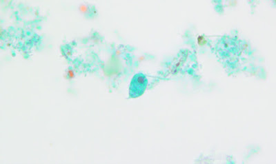
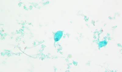
This week's case was generously donated by Drs. Jacob Rattin, Anisha Misra, and Hannah Wang, and originally identified by Marissa Roberts. The following objects were seen on a trichrome-stained stool specimen and measure approximately 15 micrometers in length. What is your identification?
Wednesday, November 1, 2023
Answer to Case 729
Answer to the Parasite Case of the Week 729: Trophozoites of Chilomastix mesnili
Below is an answer written by Dr. Jacob Rattin (@EternalStudying):
This non-pathogenic flagellate has pear-shaped trophozoites that are 6 – 24 μm long with a longitudinal spiral groove running along the body (not seen in this specimen). If visualized in a fresh prep, the motility may allow the spiral groove to be seen as the organism turns (see Case 475). The trophozoite has one nucleus (arrowhead), usually at the anterior end, with an eccentric karyosome and a cytostome (oral groove) close by. The posterior end tapers to a point (arrow).
Differentiating C. mesnili trophozoites from other non-pathogenic flagellates such as Enteromonas hominis, Pentatrichomonas hominis, and Retortamonas intestinalis can be quite difficult. Thankfully, these organisms are non-pathogenic. However, the following features may be useful in teasing apart these fun flagellates!
Enteromonas hominis trophozoites measure 5 – 7 µm long with one nucleus and three anterior flagella and one posterior flagellum. It has a single nucleus with a large karyosome. There is no cytostome which helps to differentiate it from C. mesnili.
Retortamonas intestinalis are 4 – 10 µm long with an ovoid trophozoite form with a cytostome at the anterior half that is bordered by a fibril. The single nucleus is at the anterior end and has a small karyosome.
Pentatrichomonas hominis. The trophozoites are pyriform in shape, measuring 6 – 20 µm long. They have five flagella, with four directed anteriorly and a fifth directed posteriorly. The fifth flagellum forms the outer edge of an undulating membrane and projects beyond the posterior. A single nucleus is at the anterior end and contains a small karyosome.
Thanks again to Dr. Rattin for taking on the non-pathogenic flagellates! I'm sure you'll agree that he did an excellent job. Thanks also to the laboratory technologist, Marissa Roberts, who took these photos, and to Dr. Anisha Misra at the Cleveland Clinic for her scientific oversight.