Saturday, March 25, 2017

Celebrating 10 years of Creepy Dreadful Wonderful Parasites

Thank you for joining me today to celebrate 10 years of blogging with Creepy Dreadful Wonderful Parasites! I can't believe that my very first entry on this blog was posted on March 25, 2007.

For my celebration, I invited all readers to submit their artistic parasite creations, and was amazed by all of the outstanding entries I received. They are all below for your viewing pleasure. I entered the name of each person who submitted something into a hat and then randomly selected 5 names.
And the winners are:
  • Rachael Liesman
  • Sidnei Silva
  • Prakhar Vijay
  • Melanie Bois
  • Kevin Barker
I will contact each winner separately about your prize (choice of a parasite calendar, phone case, or mug).

Fabulous parasite creative works (by category):

DRAWINGS AND PAINTINGS

Giardia and Leishmania, adorned in traditional Brazilian costumes. 
Illustrated by Sidnei da Silva

Trypanosoma brucei
Charcoal sketch by Emily Evans

Schistosoma couple
Painted on canvas by Prakhar Vijayvargiya

Tsetse fly and Reduviid bug
Illustrated by Amy Gallimore


PHOTOGRAPHY
Plasmodium berghei with a "Friday feeling Smile" by Kevin Barker

Looking for anisakids - a fish dissecting party with Rachel Vaubel, Melissa Blessing, Xuemei Wu, Emily Patterson, Melanie Bois and Heidi Lehrke

Cerebral toxoplasmosis by Melissa Blessing

MIXED MEDIA

Dipylidium caninum scarf by Heidi Lehrke

Giardia kiteii by Florida Fan

Leishmania cross-stitch by Tiffany Borbon

EDIBLE PARASITE GOODIES
Dracunculus medinensis and myiasis cupcakes by Rachael Liesman

Plasmodium falciparum cookies by Emily Fernholz

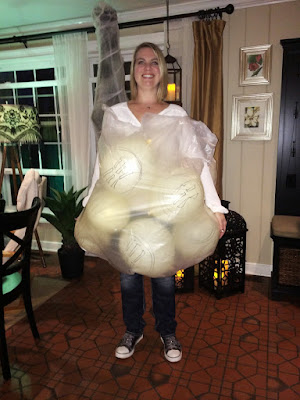
 PARASITE COSTUMES
Dermatobia hominis baby by Reeti Khare

Dipylidium caninum by Heather Rose

"Tubes of blood" for FIL (filariasis) and MAL (malaria) testing by Felicity Norris, Aimee Boeger and Brenda Nelson (note that Aimee is a green top tub (heparin) which we don't accept in my lab, so she has been 'cancelled'

Taenia solium with detachable 'proglottids' by Jadee Neff and family

Taenia solium and eosinophils with Rose Sandell and Melanie Bois

"Entamoeba histolytica/E. dispar/E. moshkovskii/E. bangladeshi and E. hartmanni - who can tell them apart?" with Patty Wright, Corrisa Miliander, Heather Rose, Emily Fernholz and Kelli Black

An engorged and gravid tick by Elli Theel

Giemsa by Jane Hata

A fish with a tapeworm by Rachael Liesman

POETRY
by Blaine Mathison
Twas the night before Christmas when all through the house
the fleas were all nestled in the fur of the mouse.
They paired with their loved ones under a sprig of mistletoe,
A gift from Cousin Chigoe, from down south in the toe.

The larvae were pupating in the bed of the host,
carrying Dipylidium cysticercoids, an infectious dose!
The Yersinia pestis churned in the foregut
until such time when the proventriculus would erupt!

All of a sudden there appeared such a clatter!
The fleas sprang from the fur to see what was the matter.
Crawling up the leg of the host, with such stealth and so quick,
was the holiday icon known as St. Tick.

“Now Ixodes, now Dermacentor, and Amblyomma!
On RhipicephalusOrnithodoros, don’t forget Hyalomma.”
He got right to work and delivered the fleas' presents
full of pathogens to spread to medieval peasants,

Then he sprang to his sleigh and let out a whistle,
Then they took off into the night like a guided missile.
But I heard him exclaim as flew out of sight,
Merry Christmas to all, and to all a good bite!

Monday, March 20, 2017

Case of the Week 439

This week's case is anticipation of Saturday, March 25th, in which I will be celebrating the 10-year anniversary of Creepy Dreadful Wonderful Parasites. As part of the celebration, I am asking all readers to consider sharing a photograph of your parasite creations - be it a painting, photograph, parasite-related costume, poem or other creative work. I will enter the name of each person who submits something in a drawing and will select 3 winners for creepy dreadful wonderful parasite prizes.

I have already received multiple entries, and have chosen to highlight this excellent entry from Florida Fan - a parasite kite! Can you guess the parasite?


Sunday, March 19, 2017

Answer to Case 439

Answer: Giardia!

This lovely kite by Florida Fan was also identified as: Giardia kitelia, Giardia flier, and Giardia lampshadia

Be sure to check out all of the fantastic parasite creations that were submitted for my 10-year celebration.

Tuesday, March 14, 2017

Celebrating 10 Years of Creepy Dreadful Wonderful Parasites - Submit your parasite works of art to be entered in a drawing!

Dear Readers,
Thank you for all of the great suggestions on how to celebrate my 10 years of blogging. After some consideration, I've decided to use this as an opportunity to appreciate the creativity of all of my readers.

As we know, parasites can be creepy and dreadful, but they can also be wonderful and beautiful in their own right. Many of you have produced delightful parasite-related creations  - original photographs, paintings, sculptures, poems, short stories, and even kites! Therefore, please consider sharing your creations with the other readers by sending me an electronic copy of your work that I can post on my blog. I will enter the name of each person who sends me something into a drawing and select 3 individuals to receive Creepy Dreadful Wonderful Parasite prizes. I will also display all of the photos/creative works that are submitted on my blog post on March 25th, 2017 - the exact date of my 10-year anniversary.

Submissions are accepted immediately! You can submit as many as you want, but your name will only be entered once. I can't wait to see all of your works of art!

Monday, March 13, 2017

Case of the Week 438

I apologize that this week's case is rather disturbing - and not in usual the creepy crawly parasite way.

The following structure was coughed up by a 70-year-old woman with advanced stage lymphoma. She had recently received chemotherapy consisting of rituximab, ifosfamide, carboplatin and etoposide (R-ICE). At the time, she was quite ill and in the intensive care unit. Likely identification?


Sunday, March 12, 2017

Looking for Ideas - Celebrating 10 years of Creepy Dreadful Wonderful Parasites

I can't believe that my first post was almost 10 years ago now - on March 25th, 2007. I feel like I have to do something special for my 10-year anniversary on 3/25/2017.

So I'm looking for ideas. Maybe a contest with prizes? I have lots of Creepy Dreadful Wonderful Parasites 'swag' to give away, including calendars, magnets, greeting cards and iPhone cases :)

Please email me with ideas.

Answer to Case 438

Answer: Sloughed esophageal mucosa.

Although this has a superficial appearance of a worm, it actually consisted of a floppy tubular structure (not fully shown in the images) composed of the outer layer of this patient's esophagus. This is a well-described phenomenon that is most commonly seen in older patients who are receiving a number of medications. It has also been seen with infectious and bullous diseases. Here are a few references which describe this phenomenon:
Carmack et al. 2009
Purdy et al. 2012
Moawad et al. 2016
Fortunately, this is usually a self-limited condition and patients fully recover.

The following images are from a similar case that was donated by Dr. Julie Ribes.

Microscopy revealed show that the structures are composed of sheets of squamous epithelial cells.


Some readers also suggested that the original case represented mucous casts, which is another good consideration. However, mucous casts would appear more solid rather than tubular.

The most important consideration in this case is not to mistake the structures for worms, since that would lead the clinical team down the wrong path in their evaluation.

Many thanks to Dr. Ribes who provided the amazing images of her similar case, and to Dr. Grys for providing the original case.

Tuesday, March 7, 2017

Case of the Week 437

This week's case was generously donated by Dr. Mike Mitchell and Janice Gotham. This is a trichrome-stained stool specimen. While the diagnosis may seem straight-forward, there is a question in what to call this organism based on some new proposed nomenclature changes. Please tell me how you think this parasite should be reported out to the patient's physician. Dr. Stensvold, I'm particularly interested in your opinion!


Monday, March 6, 2017

Answer to Case 437

Answer: Blastocystis species

I had posed the question about nomenclature to see how labs are currently reporting this parasite when it is identified in stool specimens by microscopic examination.

Originally, only B. hominis was thought to infect humans, while other species infected other animals. However, we now know that humans can be infected with the same organisms as many other hosts, and differentiation between these species is not possible by morphologic examination alone. Therefore, the most accurate way to report this organism when observed in human stool specimens is "Blastocystis sp." rather than "Blastocystis hominis".

This may cause some confusion among clinicians and therefore, providing some education about the change is recommended.

Some other fun taxonomic facts about Blastocystis spp.:
  • It was first described by Alexeev in 1911
  • It was thought to be a yeast due to its morphologic appearance
  • Later, Blastocystis was re-classified as a sporozoan protozoal parasite 
  • Blastocystis spp. is now believed to be a stramenopile - a group that includes diatoms, brown algae and mildew. The only other stramenopile to infect humans is Pythium. (HERE is a nice article in the Journal of Clinical Microbiology about human infection with P. insidiosum)
For more information about Blastocystis spp., check out the excellent BLOG by Dr. Christen Rune Stenvold. Many thanks to Dr. Graham Clark for his assistance with describing Blastocystis taxonomy is a comprehensible fashion.