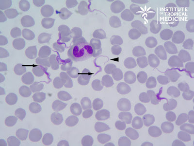
This is most likely T. brucei rhodesiense based on:
- The patient's recent travel to Eastern Africa (Kenya),
- His participation in a game tracking excursion (classic history given that wild ungulates are the reservoir for this subspecies),
- His rapid onset of symptoms, and
- The very high (!) parasitemia
Be sure to check out the video which show the characteristic 'auger' like motility of the trypomastigotes (i.e. rotating along its long axis).
Thanks to everyone who wrote in with the excellent comments. A lot of good points were raised by all. Ali Mokbel mentioned that we can't exclusively rule out T. b. gambiense, given that the patient is a frequent traveler and may have been to West Africa. Idzi also reminded us that the trypomastigotes of T. brucei are indistinguishable from those of T. rangeli, a non-pathogenic New World trypanosome which can occasionally infect humans. Fortunately, we can tentatively rule out these other species/subspecies based on the very high parasitemia and patient's symptoms. If there was any question about the identification (e.g. based on the patient's travel history), sub-species determination using PCR could be performed.
Finally, LS reminded us of the importance of determining whether the patient had central nervous system involvement since that would change the therapy. If suspected, a lumbar puncture could be performed to look for trypomastigotes.
For our students of parasitology, the following contains some general information about trypomastigotes, the most trypanosome stage seen in peripheral blood. Trypomastigotes are extracellular, unlike Plasmodium parasites, and may be seen 'swimming' between the red blood cells as in this case (check out Idzi's really cool video!). Although they have a somewhat 'worm-like' appearance, they are protozoa (not helminths), and can be easily differentiated from microfilariae by their small size (14 to 33 micrometers in length). They have a kinetoplast at their posterior end (arrows in image below) and a centrally located nucleus. A flagellum arises from the basal body (associated with the kinetoplast) and travels along the long axis of the trypomastigote as an undulating membrane. It projects from the anterior end as a free flagellum (arrow head, below), and provides the characteristic 'auger-like' motility of the trypomastigote.
Note that the flagellum is at the anterior end of the trypomastigote, and not the posterior as many would expect!
The size of the kinetoplast is very useful for differentiating the trypomastigotes of T. brucei/T. rangeli from those of T. cruzi. As you can see from the image below, the kinetoplast of T. cruzi is much larger:


1 comment:
thanks
Post a Comment