This week's case was donated by Dr. Lars Westblade. The patient is a middle-aged man who recently returned from Tanzania. He presented with multiple furuncular lesions including the following:
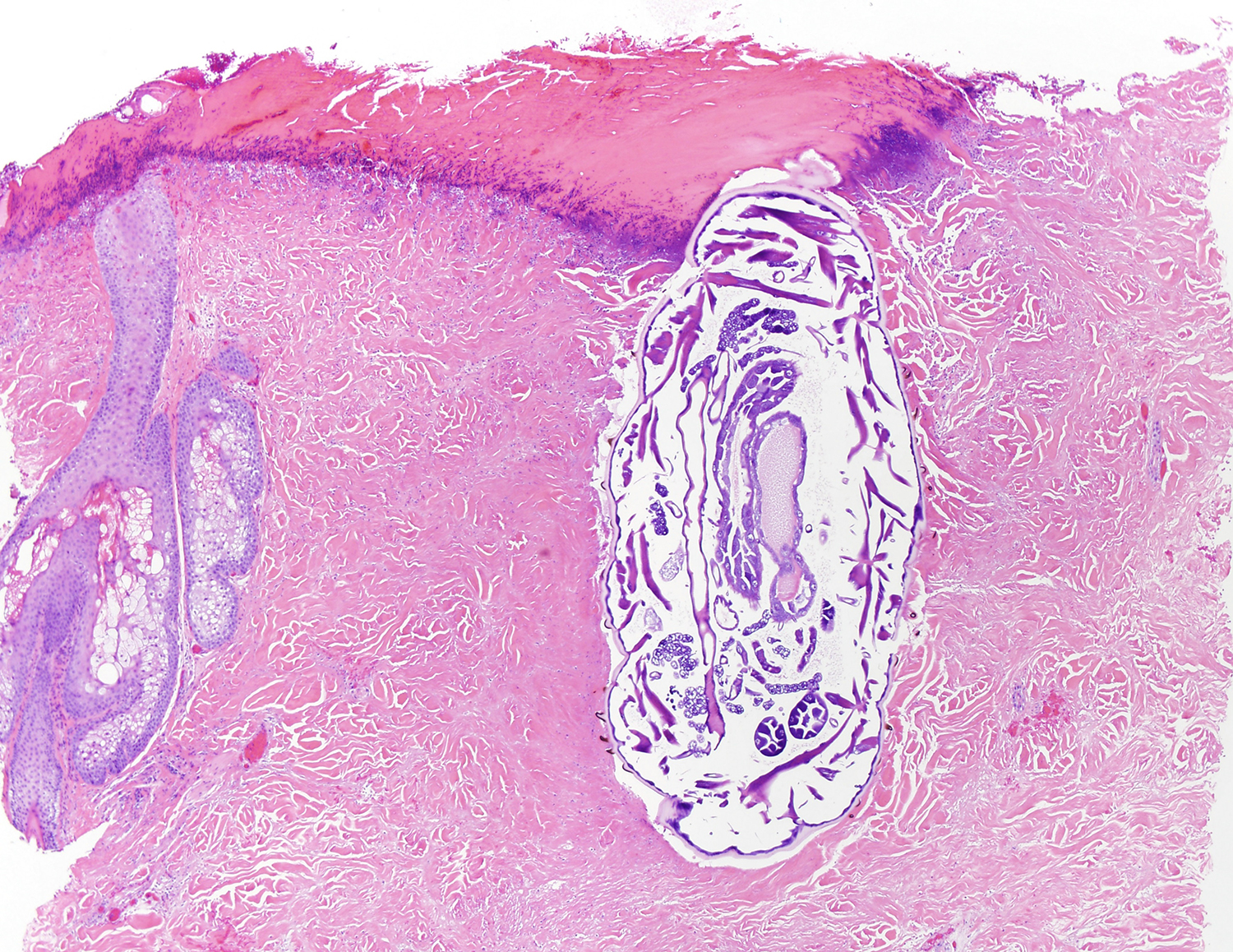
The patient saw a dermatologist who performed a skin biopsy. Here are representative sections (stained with hematoxylin and eosin):
What is the diagnosis?
13 comments:
Anonymous
said...
From the last picture, I believe we can see the mandible of the larva. Given the fact that the patient had been in Tanzania, we can narrow the diagnosis to myasis possibly caused by the tumbu fly or Cordylobia anthropophaga. Florida Fan
Indeed Florida Fan, we can see spines on the larva’s cuticle. I’d go for Cordylobia sp. (anthropophaga = Tumbu fly, or rodhaini = Lund’s fly). For differentiation I’d like to see the spiracular slits as the ones for rodhaini are sinusoidal. Strong pigmentation of the spines suggests anthropophaga though... Rodhaini also is very exceptional...
Not knowing much about entomology or histological sections, my best guess would be Cordylobia anthropophaga. This is based on the clinical presentation of furuncular lesions, the patient's travel history, and the appearance of spines on the histological section. But that is just a guess from an unexperienced parasitology enthusiast! 🙂
I agree the spiny thick cuticle and internal anatomy point to a robust dipteran larva. The furuncular lesion as well as the travel history (Tanzania) point to Cordylobia sp. Playing the geographical odds it's more likely to be C. anthropophaga.
Do these biopsies seem a it too large ?
Back in old, cold Minnesota we had 2 interesting human infestations. One was an new born infant with a 3ed stage Cuterebra bot in his neck. We were never certain how the non ambulatory baby became infested since the larvae are picked up at the entrance of animal burrows.
The second was a vet. student who had a furuncular lesion on his neck. He could see the spiracular plate moving within the lesion and actually heard a buzzing sound. He ended up at the emergency room for help. Between the noise and the pain he was being driven to the edge. He pleaded with the Dr. that there was something alive and moving in his neck. The Dr. ignored him and diagnosed his problem as psychiatric. He was put in restraints and locked up for observation. It wasn't until the Sarcophagid larvae made it's appearance that he was finally released.
In view of the clinical aspect, I think like many others that it is myasis. The existence of a multiple location is usually related to a contamination of the laundry (and the lack of ironing). The pressure of one of the lesions should have allowed to expel the larvae and to make the diagnosis on the simple clinic quickly without resorting to an expensive anatomical examination.
I agree with myasis, probably the larva of C. anthropophaga, the only definitive way to identify the species would be the posterior spiracles with 3 sinuous openings (not shown). As said before, i do not see the advantage of doing an invasive procedure as a skin biopsy when the many furuncular lesion (boil-like) and travel history points to myasis.
You're totally right Idzi. The specimen in this case could simply be a biopsy tissue that has been processed by anatomic pathology. As such, we do not have the posterior aspect for further identification details. Domage! Let's see Blaine's comments. Florida Fan
yes this is furuncular myiasis; the epidemiology supports Cordylobia anthropophaga. Cordylobia rodhani is also in Tanzania but less-commonly documented as a source of human myiasis.
Morphologic features shown in the cuts include the gut, tracheae, and striated musculature. Unlike with tungiasis, eggs are not present as this is a sexually-immature larva. The yellow spines are indeed the cuticular spices; sclerotized chitin (a carbohydrate in the arthropod exoskeleton) usually stains yellow in H&E).
Every week I will post a new Case, along with the answer to the previous case. Please feel free to write in with your answers, comments, and questions. Also check out my image archive website at http://parasitewonders.com. Enjoy!
The Fine Print: Please note that all opinions expressed here are mine and not my employer. Information provided is for educational purposes only. It is not intended as and does not substitute for medical advice. I do not accept medical consults from patients.


13 comments:
From the last picture, I believe we can see the mandible of the larva.
Given the fact that the patient had been in Tanzania, we can narrow the diagnosis to myasis possibly caused by the tumbu fly or Cordylobia anthropophaga.
Florida Fan
Actually there are spines around the body of the object and thus the diagnosis of myasis would be plausible.
Florida Fan
Credo che si tratti di Tunga penetrans.
Indeed Florida Fan, we can see spines on the larva’s cuticle.
I’d go for Cordylobia sp. (anthropophaga = Tumbu fly, or rodhaini = Lund’s fly).
For differentiation I’d like to see the spiracular slits as the ones for rodhaini are sinusoidal.
Strong pigmentation of the spines suggests anthropophaga though...
Rodhaini also is very exceptional...
I was part of the original consult on this case so I won't contribute anything yet but I can say that so far I like what I am seeing ;-)
Not knowing much about entomology or histological sections, my best guess would be Cordylobia anthropophaga. This is based on the clinical presentation of furuncular lesions, the patient's travel history, and the appearance of spines on the histological section. But that is just a guess from an unexperienced parasitology enthusiast! 🙂
I agree the spiny thick cuticle and internal anatomy point to a robust dipteran larva. The furuncular lesion as well as the travel history (Tanzania) point to Cordylobia sp. Playing the geographical odds it's more likely to be C. anthropophaga.
Do these biopsies seem a it too large ?
Back in old, cold Minnesota we had 2 interesting human infestations. One was an new born infant with a 3ed stage Cuterebra bot in his neck. We were never certain how the non ambulatory baby became infested since the larvae are picked up at the entrance of animal burrows.
The second was a vet. student who had a furuncular lesion on his neck. He could see the spiracular plate moving within the lesion and actually heard a buzzing sound. He ended up at the emergency room for help. Between the noise and the pain he was being driven to the edge. He pleaded with the Dr. that there was something alive and moving in his neck. The Dr. ignored him and diagnosed his problem as psychiatric. He was put in restraints and locked up for observation. It wasn't until the Sarcophagid larvae made it's appearance that he was finally released.
In view of the clinical aspect, I think like many others that it is myasis. The existence of a multiple location is usually related to a contamination of the laundry (and the lack of ironing). The pressure of one of the lesions should have allowed to expel the larvae and to make the diagnosis on the simple clinic quickly without resorting to an expensive anatomical examination.
I agree with myasis, probably the larva of C. anthropophaga, the only definitive way to identify the species would be the posterior spiracles with 3 sinuous openings (not shown). As said before, i do not see the advantage of doing an invasive procedure as a skin biopsy when the many furuncular lesion (boil-like) and travel history points to myasis.
You're totally right Idzi. The specimen in this case could simply be a biopsy tissue that has been processed by anatomic pathology. As such, we do not have the posterior aspect for further identification details. Domage! Let's see Blaine's comments.
Florida Fan
MYASIS
CARBUNCLE MYASIS
yes this is furuncular myiasis; the epidemiology supports Cordylobia anthropophaga. Cordylobia rodhani is also in Tanzania but less-commonly documented as a source of human myiasis.
Morphologic features shown in the cuts include the gut, tracheae, and striated musculature. Unlike with tungiasis, eggs are not present as this is a sexually-immature larva. The yellow spines are indeed the cuticular spices; sclerotized chitin (a carbohydrate in the arthropod exoskeleton) usually stains yellow in H&E).
Post a Comment