Sunday, April 30, 2023

Answer to Case 716

 Answer to the Parasite Case of the Week 716: Cyclospora cayetanensis oocysts

On the wet mount preparation with Lugol's iodine, one can nicely appreciate the spherical oocyst with well-defined outer wall and internal globular bodies. The oocysts are not highlighted by the Heine and trichrome stains (latter not shown), but their presence may be suspected by their negative contour. Confirmation can be made by staining with modified acid fast stain (or modified safranin stain), and by demonstrating autofluorescence with fluorescence microscopy. 

It is important to measure the oocysts on the stains above, since cryptosporidium oocysts have a similar appearance. C. cayetanensis oocysts are 8-10 micrometers in diameter, while Cryptosporidium spp. oocysts are 4-5.

Tuesday, April 25, 2023

Case of the Week 715

This week's interesting case was donated by Dr Mike Mitchell and his fantastic parasitology experts, Laura Derderian, Gwen Kee, and Brie Ray. The specimen below was submitted to the laboratory for evaluation of a possible parasite after being found in a 19 month girl's diaper. Identification? What is the significance to the patient?


Sunday, April 23, 2023

Answer to Case 715

 Answer to the Parasite Case of the Week 715: Cockroach ootheca (egg case or 'purse')

These fascinating structures contain about 30-40 eggs each. The female cockroach carries her ootheca until the eggs hatch and the first instar nymphs emerge. For those of you who had never heard of this phenomenon before, you can read more HERE

In this case, we can see the emerging nymphs:

Laura noted that the ootheca was hard and solid when it first arrived in the lab so she added some saline and waited for 30 minutes. This is when the nymphs began to poke out and she was able to remove them for individual viewing. Thinking about a house infested with cockroaches and their ootheca makes me shudder!

Although cockroaches are not human ectoparasites, they have important medical and economic implications. Not only are they a source of anxiety for home owners, but they also contaminate food products with their feces and secretions, cause allergic responses in susceptible individuals, and can serve as mechanical vectors and reservoirs of various pathogens such as Shigella dysenteriae, Salmonella typhimurium, poliovirus, and Entamoeba histolytica.

Thanks again to Dr. Mitchell, Laura, Gwen, and Brie for donating this great case! 

Tuesday, April 4, 2023

Case of the Week 714

This week's case was donated by Dr. Sandeep T. You may remember the amazing case of fasciolopiasis he provided back in 2012 (and if not, you should definitely check it out HERE). His latest case is from a middle aged taxi driver who reports passing white objects in is his stool for the past 2 years. He denies fever, weight loss, vomiting and diarrhea. A routine complete count is unremarkable with no anemia. Below is one of the white objects submitted for pathologic examination. Identification? What additional procedures would be helpful in this case?


Sunday, April 2, 2023

Answer to Case 714

 Answer to the Parasite Case of the Week 714: Taenia species proglottid. Examination of the uterine branching pattern is required for species level identification when using morphology alone. Gravid proglottids (which we know we have here since we see eggs) can be categorized into 2 groups: Taenia solium proglottids have 7-13 primary lateral branches off of the central stem, whereas T. saginata and T. asiatica have 12-30. Of the three species, only T. solium causes human cysticercosis, so it is helpful to identify when T. solium is present.

Options for determining the species of gravid proglottids are:

1. Transilluminating the proglottid to observe the uterine branching pattern. You can see a great example of this in Idzi's previous case from 2017

2. Sometimes it helps to press the proglottid between 2 slides to better visualize the branches, as seen in Idzi's case from 2018:


3. When the proglottid has been fixed in formalin or ethanol, it is sometimes necessary to clear the proglottid in lactophenol. Injecting the central uterine canal and branches with India ink, or fixing the proglottid and staining it with carmine may also be helpful. Here's a carmine-stained proglottid from back in 2015:
We don't actually use the India ink injection method in my lab, since it's rather finicky and it exposes my techs to the Taenia eggs. The eggs of T. solium are infectious to humans, so I prefer to pursue other routes to identification when possible. That leads me to the last thing that we try:
4. Histologic processing of the proglottid, with sectioning to see the uterine branches. Note that the proglottid has to be embedded just right in order to see the branches. Here are a couple of cases in which this worked out quite nicely:


Sandeep is going to section deeper into the block to see if he can appreciate the uterine branches. For now, however, we can see the confirmatory features of a Taenia sp. proglottid, with the round-to-oval eggs. 


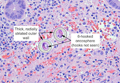
The thick wall is apparent, although the striations are not. However, I've seen many other cases that have allowed me to have an appreciation of what we are looking for. Here are some good examples from other cases:

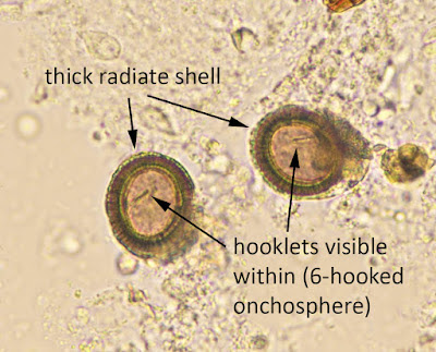
See how they compare to the typical eggs that we see in stool wet preparations:

Note that some claim that the shape of the eggs (round vs. oval) or staining with periodic acid Schiff (PAS) allows for species-level ID. However, the studies show that this is not reliable, as there is significant overlap between the two. 
5. Nucleic acid amplification tests such as PCR can also be quite helpful when available. Testing may be available at no cost through some specialty academic medical centers and the Parasite World Bank (As per Dongmin Less, you can email admin@parasiteworldbank.org). 

6. Last but not least, you can do as Lamia Galal recommends, and ask the patient what type of uncooked meat he ate!

Thank you all for the great comments and information.