Answer to the Parasite Case of the Week 714: Taenia species proglottid. Examination of the uterine branching pattern is required for species level identification when using morphology alone. Gravid proglottids (which we know we have here since we see eggs) can be categorized into 2 groups: Taenia solium proglottids have 7-13 primary lateral branches off of the central stem, whereas T. saginata and T. asiatica have 12-30. Of the three species, only T. solium causes human cysticercosis, so it is helpful to identify when T. solium is present.
Options for determining the species of gravid proglottids are:
1. Transilluminating the proglottid to observe the uterine branching pattern. You can see a great example of this in Idzi's previous case from 2017:
2. Sometimes it helps to press the proglottid between 2 slides to better visualize the branches, as seen in
Idzi's case from 2018:
3. When the proglottid has been fixed in formalin or ethanol, it is sometimes necessary to clear the proglottid in lactophenol. Injecting the central uterine canal and branches with India ink, or fixing the proglottid and staining it with carmine may also be helpful. Here's a
carmine-stained proglottid from back in 2015:
We don't actually use the India ink injection method in my lab, since it's rather finicky and it exposes my techs to the Taenia eggs. The eggs of T. solium are infectious to humans, so I prefer to pursue other routes to identification when possible. That leads me to the last thing that we try:
4. Histologic processing of the proglottid, with sectioning to see the uterine branches. Note that the proglottid has to be embedded just right in order to see the branches. Here are a couple of cases in which this worked out quite nicely:
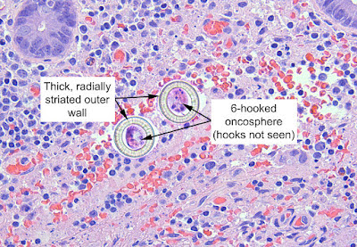
Sandeep is going to section deeper into the block to see if he can appreciate the uterine branches. For now, however, we can see the confirmatory features of a Taenia sp. proglottid, with the round-to-oval eggs.
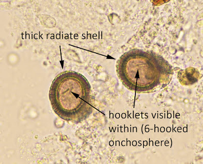
The thick wall is apparent, although the striations are not. However, I've seen many other cases that have allowed me to have an appreciation of what we are looking for. Here are some good examples from other cases:
See how they compare to the typical eggs that we see in stool wet preparations:
Note that some claim that the shape of the eggs (round vs. oval) or staining with periodic acid Schiff (PAS) allows for species-level ID. However, the studies show that this is not reliable, as there is significant overlap between the two.
5. Nucleic acid amplification tests such as PCR can also be quite helpful when available. Testing may be available at no cost through some specialty academic medical centers and the Parasite World Bank (As per Dongmin Less, you can email
admin@parasiteworldbank.org).
6. Last but not least, you can do as Lamia Galal recommends, and ask the patient what type of uncooked meat he ate!
Thank you all for the great comments and information.


2 comments:
well said ...thank you
Few additional points to add:
1. Counting of uterine branches is only reliable on gravid proglottids. Sometime you may not get a mature/gravid proglottid where there may be an overlap > 12-14 branches if there are multiple proglottids presented, try and count 2-3.
2. Although epidemiology is important, be aware that in many developing countries mincemeat
may be ‘mixed’ (pork meat mixed with beef or lamb) so just the history may not always be reliable.
3. PCR is most reliable for speciation but is not cheap. Commercial multiplex assays are available.
Post a Comment