Answer to Parasite Case of the Week 739: Loa loa microfilariae
Thanks to everyone who wrote in with comments. We received a lot of different responses including some of the sheathed and unsheathed microfilariae. Therefore, this is a great time to review my approach to identifying microfilariae in blood specimens. You can also read this article I wrote with Blaine Mathison and Marc Couturier that provides a diagnostic algorithm for microfilariae in blood. In this algorithm, we recommend first measuring the length of the microfilariae. If they are small (<200 micrometers long), then it is likely to be one of the Mansonella species. Mansonella are not sheathed, small, and quite narrow. Their width is smaller than the diameter of a neutrophil or eosinophil. If, on the other hand, the microfilariae are relatively large (>200 micrometers), then you are dealing with Wuchereria bancrofti, Loa loa, or one of the Brugia species. These are sheathed microfilariae, but the sheath may not always be readily visible on Giemsa stain. Therefore, the size is a more reliable diagnostic feature. To better visualize the sheath, a hematoxylin stain can be performed such as the Carazzi or Delafield hematoxylin stain. (Note: the hematoxylin and eosin stain used in histopathology preparations can also be used, so if you don't have a hematoxylin stain for microfilariae, you can ask for help from your friends in anatomic pathology!) These larger microfilariae can then be differentiated by characteristics of their head and tail spaces.
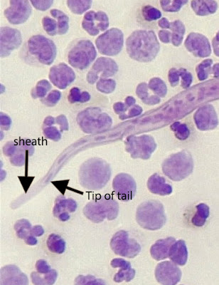
With that introduction, let's turn to the specifics of this week's case. I forgot to provide the length - my apologies! - I've now added it to the case description (~250 micrometers long). You can also get a general sense of the size of the microfilariae by comparing their diameter to the surrounding white blood cells. Note in this image below that the width of sheathed microfilariae such as Loa loa is slightly greater than the diameter of the neighboring eosinophils. Also, there is a hint of sheath (arrows). In contrast, the Mansonella perstans microfilaria in the image on the right is narrower than the surrounding eosinophils.
Importantly, you can also see a sheath with the Carazzi stain that Idzi provided. Therefore, we can rule out the two Mansonella species found in blood by this finding alone.
Note that the nuclei go to the tip of the tail, which makes this a classic case of Loa loa. (one of my favorite memory aids is that the nuclei 'flow-a flow-a to the tip in Loa loa).
To answer my second question - the additional study that should be considered in this case is calculation of the microfilarial burden since patients with >8,000 microfilariae/mL are at risk of encephalopathy when treated with diethylcarbamazine (DEC).
Thanks again to Idzi Potters and the Institute of Tropical Medicine, Antwerp, for this great case! The next case will come out early next week.


No comments:
Post a Comment