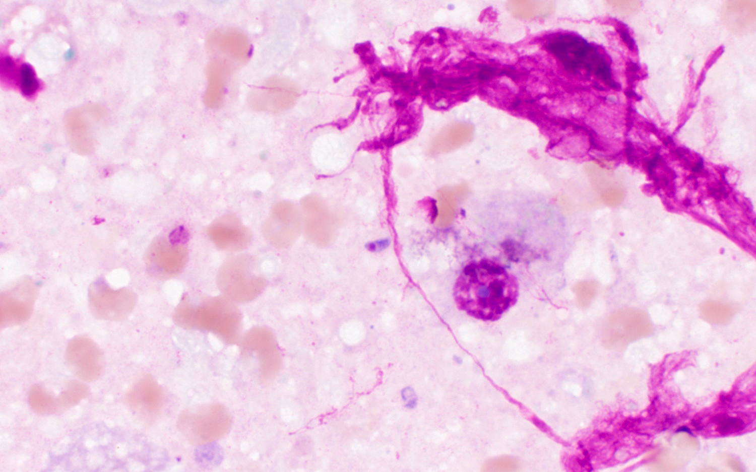
This week's case is a brain biopsy from a middle-aged man with untreated HIV. The specimen appeared necrotic and bloody. Touch preps were made from the material and stained with Giemsa. From the images below taken with the 100x oil objective, what is your diagnosis?


3 comments:
It looks like Toxoplasma gondii (and that would make sense to be actively replicating with HIV). Looks like free and intracellular tachyzoites
I would agree with the previous answer. There are free tachyzoites, intracellular ones and I believe there is a cyst also. Usually these cases are referred to the Pathology department and most Parasitology staff don’t have a chance to see them.
I am thinking Microsporidia, they usaully seen in clumps or in clusters
Post a Comment