Monday, September 19, 2011

Case of the Week 176

The following objects were seen in cross-section in an appendix from a 14 year old girl with abdominal pain. Identification? (CLICK ON IMAGES TO ENLARGE)

(H&E, 40x original magnification)


(H&E, 100x original magnification)


(H&E, 200x original magnification)

(H&E, 40x original magnification)


(H&E, 100x original magnification)


(H&E, 200x original magnification)

Sunday, September 18, 2011

Answer to Case 176

Answer: Enterobius vermicularis (pinworm) cross-sections

This is a classic histologic appearance of E. vermicularis in cross-section. The diagnosis is made by identification of the lateral alae (singular=ala, from the Latin meaning "wing"). Indeed, you can see why these longitudinal ridges on either side of this roundworm's body have been likened to wings.

Other features include well-developed platymyarian-type musculature (Body musculature consisting of cells not protruding individually into Pseudocoelom), thick muscled esophagus, and thick nucleated intestine; features seen in many intestinal nematodes (round worms).

Some of these features are shown in Case of the Week 88

Thanks to everyone for writing in!

Sunday, September 11, 2011

Case of the Week 175

The following objects were seen in unstained fluid aspirated from a liver cyst and sent to microbiology. The beautiful images were taken by Heather A. Identification?


Saturday, September 10, 2011

Answer to Case 175

Answer: Echinococcus spp. protoscoleces

Shown here are multiple protoscoleces (larval tapeworms) of Echinococcus spp. The most common species to infect humans is Echinococcus granulosus, but E. multilocularis and less commonly, other species, may also cause human infection. Note that one larva has an everted protoscolex (below).


Based on the appearance of the everted protoscolex (above), you can see how this larval form will mature into the adult Echinococcus worm in the definitive canid host:

Adult Echinococcus granulosus worm


Higher magnification reveals the hooklets of the scolex, used to attach to the intestinal tract of the definitive host.


In echinococcosis, humans and herbivores are the intermediate host and are infected with the larval forms, whereas members of the Canidae such as wolves and domestic dogs are the definitive hosts and are infected with adult tapeworms.

Monday, August 29, 2011

Case of the Week 174

The following were found in a vacant ski lodge which had not been occupied in several months. Identification? (CLICK ON IMAGE TO ENLARGE)


Sunday, August 28, 2011

Answer to Case 174

Answer: Bat bugs

Congratulations to all of my readers who got this right! I thought this might be a tricky case since bat bugs share many morphologic similarities with the ever-popular bed bugs. But most of you picked up on the subtle differences between the 2 arthropods.

As Blaine points out, "Given the length of the pronotal setae, they are bat bugs, Cimex sp. Cannot differentiate further without more geographic location (C. adjunctus in the east; C. pilosellus in the west)." Here are some web sites that highlight the important differences between these 2 bugs: BUGGUIDE and BADBEDBUGS

Monday, August 22, 2011

Case of the Week 173

The following sections are from a skin biopsy of an itchy flank rash. Identification?

(H&E, 40x original magnification)


H&E, 400x original magnification


H&E, 400x original magnification

Sunday, August 21, 2011

Answer to Case 173

Answer: Scabies caused by the microscopic mite Sarcoptes scabiei.

This mite burrows into the superficial layers of the epidermis and deposits eggs and fecal pellets (scybala). The diagnostic features of this case are:
1. The location within the epidermis
2. Size of mite and presence of toothlike spines on its thin exoskeleton (see below).
3. Adult and egg forms (below)


The best way to diagnose scapies is via skin scrapings (rather than biopsy). Skin scrapings can be obtained through which will produce mites with the best morphology. To obtain skin scrapings, mineral oil can be applied to the possible skin tracts followed by vigorous scraping with a sterile scalpel blade or glass slide. The material obtained should be placed on glass slides and examined for the presence of mites, larvae, and fecal pellets.

Anonymous and Neuronurse mentioned that this parasite resembles Gnathostoma spp, which also have spines on their external surface. However, Gnathostoma are uncommon in the epidermis (usually seen in subcutaneous tissue and viscera) and the spines are much smaller than those seen on the scabies mite. Gnathostoma also has different internal structures including large lateral cords. Good thought!

Monday, August 15, 2011

Case of the Week 172

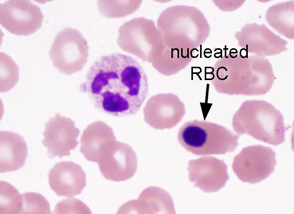
A 35 year old immigrant from Sudan presents with fever. Images from a Giemsa-stained thin blood film are shown. Diagnosis? (CLICK ON IMAGES TO ENLARGE)


Sunday, August 14, 2011

Answer to Case 172

Answer: No blood parasites present; Howell-Jolly bodies, platelets, and a nucleated RBC present).

Great job everyone! Several respondents rightly raise the question of asplenia, given the Howell-Jolly bodies (nuclear remnants), nucleated RBCs, and RBC polymorphism with echinocytes (wrinkled RBCs with multiple spicules radiating from its circumference, resembling a sea urchin). These are common findings in a patient that does not have a functional spleen, since the spleen is responsible for removing RBCs that have not fully extruded their nucleus prior to entering the peripheral blood. We found out that this patient does NOT have a spleen which would explain the findings on this film.


This is a great example of a 'complicated' peripheral blood smear, in which
the exclusion of malaria or babesiosis can be quite difficult. The presence of Howell-Jolly does not exclude the presence of a blood parasite, so it is still important to examine these smears carefully.
Anonymous raises the good point that PCR for malaria and babesiosis may be helpful when either of these infections are suspected. In this case, there was no evidence of blood parasites seen on thick and thin films, so we did not pursue PCR.

Other types of RBC "inclusions" include pappenheimer bodies (iron granules) and stain precipitate.

Finally,
Anonymous mentions that the "tiny intraerythrocytic inclusions may be bacterial, like Bartonella. B. bacilliformis is the only one that's supposed to show up on peripheral smear, but no epidemiologic history to fit." This is a good thought, although as mentioned, it does not fit clinically. I would also expect the inclusions of B. bacilliformis to be more numerous and smaller than what is shown here.