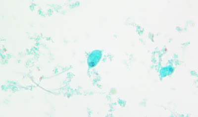
This week's case was generously donated by Drs. Jacob Rattin, Anisha Misra, and Hannah Wang, and originally identified by Marissa Roberts. The following objects were seen on a trichrome-stained stool specimen and measure approximately 15 micrometers in length. What is your identification?
Thursday, November 2, 2023
Wednesday, November 1, 2023
Answer to Case 729
Answer to the Parasite Case of the Week 729: Trophozoites of Chilomastix mesnili
Below is an answer written by Dr. Jacob Rattin (@EternalStudying):
This non-pathogenic flagellate has pear-shaped trophozoites that are 6 – 24 μm long with a longitudinal spiral groove running along the body (not seen in this specimen). If visualized in a fresh prep, the motility may allow the spiral groove to be seen as the organism turns (see Case 475). The trophozoite has one nucleus (arrowhead), usually at the anterior end, with an eccentric karyosome and a cytostome (oral groove) close by. The posterior end tapers to a point (arrow).
Differentiating C. mesnili trophozoites from other non-pathogenic flagellates such as Enteromonas hominis, Pentatrichomonas hominis, and Retortamonas intestinalis can be quite difficult. Thankfully, these organisms are non-pathogenic. However, the following features may be useful in teasing apart these fun flagellates!
Enteromonas hominis trophozoites measure 5 – 7 µm long with one nucleus and three anterior flagella and one posterior flagellum. It has a single nucleus with a large karyosome. There is no cytostome which helps to differentiate it from C. mesnili.
Retortamonas intestinalis are 4 – 10 µm long with an ovoid trophozoite form with a cytostome at the anterior half that is bordered by a fibril. The single nucleus is at the anterior end and has a small karyosome.
Pentatrichomonas hominis. The trophozoites are pyriform in shape, measuring 6 – 20 µm long. They have five flagella, with four directed anteriorly and a fifth directed posteriorly. The fifth flagellum forms the outer edge of an undulating membrane and projects beyond the posterior. A single nucleus is at the anterior end and contains a small karyosome.
Thanks again to Dr. Rattin for taking on the non-pathogenic flagellates! I'm sure you'll agree that he did an excellent job. Thanks also to the laboratory technologist, Marissa Roberts, who took these photos, and to Dr. Anisha Misra at the Cleveland Clinic for her scientific oversight.
Wednesday, October 11, 2023
Case of the Week 728
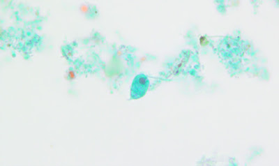
This week's case was donated by Drs. Meredith Kavalier, Megan Shaughnessy, and David Cartwright. The patient has a history of treated urothelial cell carcinoma and remote travel to Asia. The cytopathologist was concerned by the presence of the following structures seen in a routine screening urine sample. How would you interpret these findings?
Tuesday, October 10, 2023
Answer to Case 728
Answer to the Parasite Case of the Week 728: Not a parasite egg; uric acid crystals.
Uric acid crystals are a very convincing mimic of Schistosoma haematobium eggs! They are both found in urine, and they both have a terminal spine. However, there are a number of features that can be used to easily differentiate the two:
- Uric acid crystals vary in size and shape and are often much smaller than S. haematobium eggs. In contrast, S. haematobium eggs are regular in size and shape, and quite large (approximately 150 micrometers in length).
- Uric acid crystals commonly have points on both ends instead of the single 'pinched-off' spine of S. haematobium eggs. They can also have lateral points or take on other shapes.
- There are no internal parasite structures (i.e., miracidium) in uric acid crystals
- Finally, crystals often fracture and break, and may have irregular contours.
Tuesday, September 26, 2023
Case of the Week 727
his week's case was generously donated by Dr. Richard Bradbury. The following structure was seen in a concentrated wet prep of stool. It measures approximately 80 micrometers in diameter. Check out the video to see this structure in multiple planes. Identification?
Monday, September 25, 2023
Answer to Case 727
Answer to the Parasite Case of the Week 727: Hymenolepis diminuta
As noted by Lamia Gala on LinkedIn, "The distance between the outer and the inner shell is large with no polar filaments" which is consistent with H. diminuta. This is in contrast to Rodentolepis (formerly Hymenolepis) nana, which does have polar filaments between the outer and inner shell. H. diminuta is also slightly larger than R. nana, measuring 70-85 micrometers in greatest dimension (compared to 30-50 for R. nana). Florida Fan provided the helpful memory aid that the word "diminuta" is larger in size than "nana", which also applies to their eggs (thanks FF!). Here are some of the key morphologic features:
Thanks again to Dr. Bradbury for donating this lovely case!Monday, September 18, 2023
Case of the Week 726
This weeks case features the following small, oval-shaped, red-staining objects seen in a bronchoalveolar specimen from an immunocompromised patient. The objects measure approximately 2 micrometers long. The stain is a strong trichrome (chromatrope 2R method). Identification? What is the significance of this finding? (CLICK ON IMAGES TO ENLARGE)
Sunday, September 17, 2023
Answer to Case 726
Answer to the Parasite Case of the Week 726: Microsporidia spores; genus and species not determined
These small spores stain deep red with the chromotrope 2R method and its modifications. They are oval shaped and often have a darker-staining equatorial band in the middle of the spore. Spores of Enterocytozoon bieneusi are very small (0.8 to 1.4 micrometers long), whereas other species such as Encephalitozoon spp., Vittaforma cornea, and Anncaliia algerae are a little larger. Some (e.g., Anncaliia) are up to 4 micrometers long and may be mistaken for small years such as Histoplasma capsulatum (especially because they are focally GMS positive!)
As Dr. Couturier pointed out, microsporidia are not parasites, but fungi (his words: "Not a parasite LOL Get those buggers off this page and send them to the Mycology lab. 😉) . They are obligate intracellular, unicellular, spore-forming eukaryotic organisms comprising more than 220 genera and 1,700 species. Microsporidia itself is NOT a genus and should not be italicized.Tuesday, September 5, 2023
Case of the Week 725
This week's interesting case is generously donated by Dr. Justin Juskewitch. Hopefully all of you have been able to avoid this so far this summer!
The patient is a young girl who developed this very itchy rash about 30 minutes after a swim in a fresh water lake in Maine (Northeastern United States). The rash developed to what is shown below over a period of several hours.
Her two siblings had a similar presentation. All three children had resolution of itching with benadryl, corticosteroid cream, and oatmeal body wash baths over the next few hours, but the rash lasted for 5-7 days.
What is the most likely diagnosis? Is any additional therapy recommended?
Sunday, September 3, 2023
Answer to Case 725
Answer to Parasite Case of the Week 725: Swimmer's itch
As noted by Florida Fan, this is "typical swimmer’s itch, also known by other names depending on the activity of the patient like 'clam digger’s itch' or 'duck itch'." (Also called Pelican itch in Australia) "All are caused by [zoonotic] cercariae in most freshwater bodies of water frequented by ducks and/or water birds. The cercariae penetrate the skin and cause a cercarial dermatitis. This summer is so hot even in the Northern most states that a quick plunge into the lake is certainly very appealing. We may expect to see more cases like this one."
Indeed, this is a distressing result from what would otherwise be a lovely dip in a cool lake!
An anonymous reader elaborated that "Swimmer's itch is an allergic condition that occurs when trematode cercariae, the motile and infectious stage of avian schistosomes (eg, Trichobilharzia spp.), penetrate the skin of humans. They utilize a variety of different species of birds as definitive hosts (humans are not suitable hosts), and rely on different snail species as intermediate hosts. If any, only symptomatic therapy is needed (anti-itch lotions, antihistamine), the cercariae die quickly without causing a severe trematode infection."
Marc Couturier noted "Growing up in Maine, we just called that...the result of swimming in Maine". Yikes! It's also common in northern Minnesota and Wisconsin in the United States.
I had to laugh at the Twitter (X) response from That Packer Girl 🏈 (@thatpackergirl) - "*sideyes the mergansers and snails at the family cabin*"
In the United States, swimmer's itch 'season' is mid summer. According to a post from Anoka County, Minnesota, Parks and Recreation, you can "Reduce your chances of getting severe swimmers’ itch by following these simple guidelines. Dry off as soon as you leave the water. Rub skin briskly to remove water drops before they evaporate. Be sure to dry underneath waistbands and around leg openings. Encourage children to dry off thoroughly each time they leave the water. Shower with soap and fresh water or change into dry clothes as soon as possible. Don’t wade or play in shallow water. Swimming from a raft or pontoon minimizes your exposure. Don’t feed geese and ducks near your beach. Waterfowl are an important adult host for the parasites."
I hope you all enjoy the rest of the summer!