Answer to the Parasite Case of the Week 766: Schistosoma haematobium eggs
The following outstanding discussion is written by our guest author, Dr. Azra Hasan.
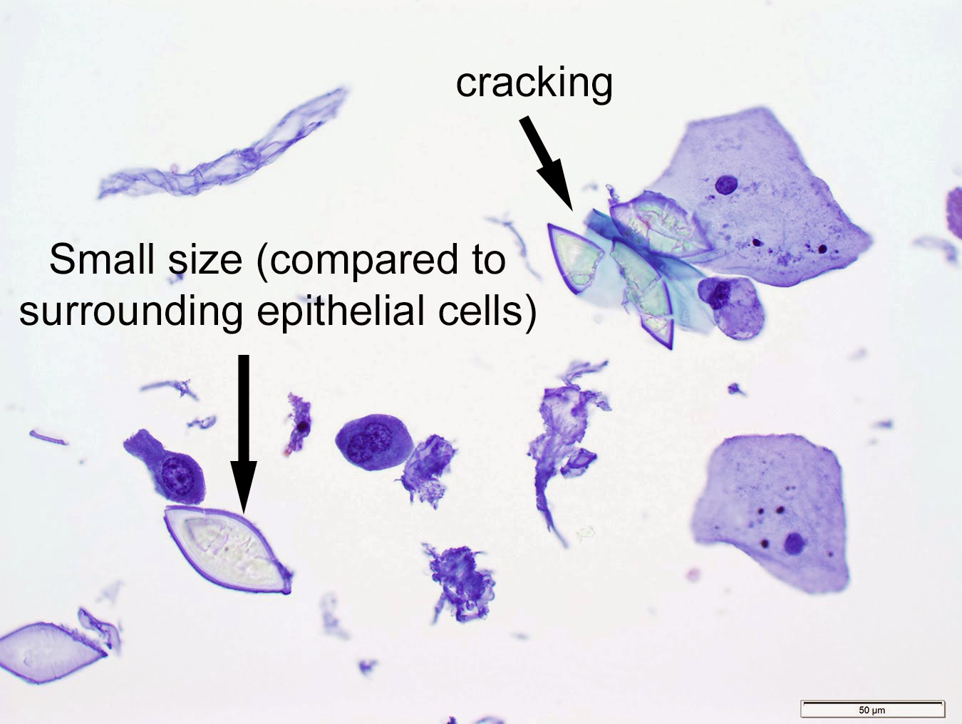
These are Schistosoma haematobium eggs in a urine sample as many of you correctly identified in the comments. The key giveaway was the prominent “terminal spine”, that helps differentiate S. haematobium from other Schistosoma species. On a closer look we can see that the miracidium (larval stage) fills up the egg. (Be sure to check out case 467 which has incredible videos from Idzi Potters, showing a beautiful motile miracidium!). Note that observing motile miracidia within eggs or in urine serves as a direct indication of an active S. haematobium infection and should be reported.

As noted by Florida Fan, typically, Schistosoma haematobium eggs are found in urine, which is a good way to differentiate them from the similar-appearing, but larger, S. intercalatum, which are found in stool. However, in heavy infections, and especially from residents of endemic areas, one may encounter S. haematobium eggs in stool as well. Rarely, they can also be seen in semen and in biopsy tissues from the bladder, as well as the genitalia and rectum. In endemic regions, nearly 75% women with urinary schistosomiasis have S. haematobium eggs in the genitalia. Female genital schistosomiasis (FGS) is often associated with eggs in the cervix, vagina, and/or vulva, with sandy patch lesions and mucosal bleeding sometimes without hematuria. (2)
Satoshi rightly pointed out the RBCs in the background indicating hematuria - During acute infections, hematuria is an important finding along with history that often includes dysuria and frequent urination. Advanced infections may lead to fibrosis, calcification, obstructive uropathy, polyps, or squamous cell carcinoma of the bladder. Imaging often reveals calcifications described as the "eggshell bladder”.
Praziquantel remains the gold standard drug for treatment, but reinfections are common. Many vaccine candidates show promise and would be the answer in endemic regions. (4)
Here are the primary components of the Schistosoma lifecycle:
- Man is the definitive host who acquires infection by free-swimming cercariae in contaminated freshwater. These penetrate the skin and advance within dermal veins to become the next stage larva. They then move to lungs, systemic circulation, portal circulation, finally developing into adults in the liver.
- Paired adults male and female worms travel to the vesical and pelvic venous plexus, anchor themselves in the venules surrounding the bladder wall and release eggs. (Scroll through Case 522 to read the eternal love story of the male and female Schistosoma!)
- To continue its lifecycle, the eggs penetrate the bladder wall (remember the terminal spine here!) and are excreted in the urine. And while this happens, some eggs get trapped in the bladder tissue. The eggs that are excreted in the urine, hatch into miracidia upon reaching fresh water and seek out their next host – the Bulinus snails.
- Bulinus snails are the intermediate hosts - Miracidia develop asexually into sporocysts which transform into free-swimming cercariae. These are released in fresh water and the cycle continues.
The following are some helpful diagnostic approaches:
- Use of reagent strips to detect hematuria and proteinuria in areas of endemic infection has proven to be an effective screening technique for S. haematobium infections.
- Eosinophilia has also been used to diagnose infections,
- S. haematobium eggs are usually detected in the urine. The terminal hematuria portion of the urine specimen may contain numerous eggs trapped in the mucus and pus. Tissue biopsy, vaginal lavage, stool, semen microscopy can be done with clinical suspicion and travel history from endemic regions. (1)
- An antigen-capture ELISA utilizing monoclonal antibodies (MAbs) such as 290-2E6-A and 128C3/3/21 is one of the most promising assays for immunodiagnosis of S. haematobium infections. Antibody detection have not been particularly useful because of cross-reactions with other helminth infections. (5)
Did you know?
- Peak egg excretion occurs between 10am and 2 p.m. Samples collected during this time, or during a 24-h urine collection without preservatives, may be used for examination. It is important to use saline and not water for the urine concentration procedures; this will avoid hatching of the eggs. (Ref: Centre for Disease Control and Prevention (CDC). Schistosomiasis - Diagnostic procedures.)
- The “fetal head” sign is seen in abdominal X rays and is caused by calcium deposits around schistosome eggs in the bladder and uterine walls
- Symptoms are usually not seen for 3 to 6 months or more. This is why S. haematobium infection is listed as a “delayed” cause of hematuria. (3)
- Hematuria is so common that in some areas of endemic infection this phenomenon in boys was considered to be analogous to menarche in girls.
- S. haematobium infection is endemic in Africa including Caribbean Islands (West Indies), Madagascar, Arabian Peninsula and one limited focus in Maharashtra, India.
Some interesting case reports and reviews:
- Case Report: A Child with Gross Hematuria and the Importance of Travel History. Shatat IF. Front Pediatr. 2018 Feb 5; 6:14. doi: 10.3389/fped.2018.00014. PMID: 29459889; PMCID: PMC5807655.
- Female genital schistosomiasis is a neglected public health problem in Tanzania: Evidence from a scoping review. Mbwanji G, Mazigo HD, Maganga JK, Downs JA. PLoS Negl Trop Dis. 2024 Mar 11;18(3): e0011954. doi: 10.1371/journal.pntd.0011954. PMID: 38466660; PMCID: PMC10927128.
- Schistosoma haematobium: A Delayed Cause of Hematuria. Tan WP, Hwang T, Park JW, Elterman L. Urology. 2017 Sep;107: e7-e8. doi: 10.1016/j.urology.2017.06.021. Epub 2017 Jun 23. PMID: 28652164.
- A comprehensive and critical overview of schistosomiasis vaccine candidates. Al-Naseri A, Al-Absi S, El Ridi R, Mahana N. J Parasit Dis. 2021 Jun;45(2):557-580. doi: 10.1007/s12639-021-01387-w. Epub 2021 Apr 25. PMID: 33935395; PMCID: PMC8068781.
- Detection of circulating antigens in patients with active Schistosoma haematobium infection. Hassan MM, Medhat A, Makhlouf MM, et al The American journal of tropical medicine and hygiene Am J Trop Med Hyg Am. J. Trop. Med. Hyg. 1998;59(2):295
More information
- CDC (Centers for Disease Control and Prevention): Schistosomiasis - Parasites (https://www.cdc.gov/parasites/schistosomiasis/)
- WHO (World Health Organization): Schistosomiasis fact sheet (https://www.who.int/news-room/fact-sheets/detail/schistosomiasis)
- London School of Hygiene & Tropical Medicine: Schistosomiasis Resources and Research (https://www.lshtm.ac.uk/research/research-action/global-research-portfolio/schistosomiasis)
- NIH (National Institutes of Health): Schistosomiasis - MedlinePlus (https://medlineplus.gov/schistosomiasis.html)



+and+crystal+(right).jpg)


