This week's case was generously donated by Dr. Jacob Rattin. The following objects were seen from Wright-stained thin blood films taken from a middle-aged woman with unexplained fevers. What is your diagnosis?


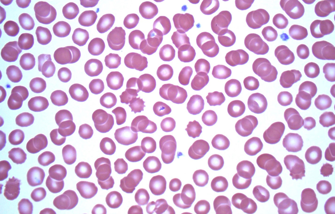
This week's case was generously donated by Dr. Jacob Rattin. The following objects were seen from Wright-stained thin blood films taken from a middle-aged woman with unexplained fevers. What is your diagnosis?
Answer to Parasite Case of the Week 754: Platelets overlying RBCs, an intraerythrocytic blood parasite mimic. Many thanks to Dr. Jacob Rattin who donated this case and provided the following helpful explanation.
Normal platelets are 1.5-2.5 μm in diameter and are morphologically round-to-oval and granular (Figure 1, arrowheads).

On Wright stain they are usually blue/purple and on Giemsa stain they are normally pink/purple. As seen in Figures 1 through 4 (arrows), platelets can overly red blood cells (RBCs), mimicking a blood parasite (e.g., Babesia or Plasmodium). In Figure 5, the green arrow is pointing to stain precipitate overlying an RBC.


Helpful morphologic clues to differentiate this pitfall from a true parasite are the following:
1. Platelets overlying RBCs often have a “halo” around them which is likely from it pressing down upon the RBC, which signifies it is not actually intraerythrocytic.
2. The platelets lack any definitive morphology of a parasite.
3. Other normal platelets in the field should resemble the platelet/s overlying the RBC (which is apparent in Figures 1 through 5).
4. As always, clinical information integration is crucial! Checking the patient’s medical and travel history can aid in the differential diagnosis.
This week's interesting case was donated by Dr. Hugh Mortan and Jodie Smith. They noted the following object in a Gram-stained bronchial lavage specimen. What is your diagnosis?


Answer to the Parasite Case of the Week 753: Not a parasite; Curshmann spirals.
As noted by several of my readers, Curshmann spirals are spiral-shaped mucus plugs from subepithelial mucous gland ducts of bronchi that occur in a variety of diseases including asthma and bronchitis.

As I always say about mimics - it's not so important that you know the name of the mimic, as long as you recognize it is not a parasite!
Florida Fan has a really good saying to help with this differentiation: "No head, no tail, no entrails, most likely no parasite”.
Thanks again to Dr. Hugh Mortan and Jodie Smith for donating this great case!


Answer to the Parasite Case of the Week 752: Botfly larva, likely Cuterebra species.
Thanks to Michele Calatri, Kate Geralt, Pablo David Jimenez Castro, Christina, and Josué Campos Camacho who wrote in with this answer. Cuterebra infection is not very common in humans but are known to occur occasionally. Unfortunately, we don't have the posterior spiracles to confirm the genus, and we don't have any further history about the patient. The good news is that the larva has been removed, which is curative.
This is how the larvae appear in their natural host:
Creepy Dreadful Wonderful Parasites: Case of the Week 130 (parasitewonders.blogspot.com
This week's case was generously donated by Dr. Karra Jones. A patient presented with intestinal intussusception, and the following was seen on histopathologic examination of the resected bowel. What is your diagnosis?
10x objective:
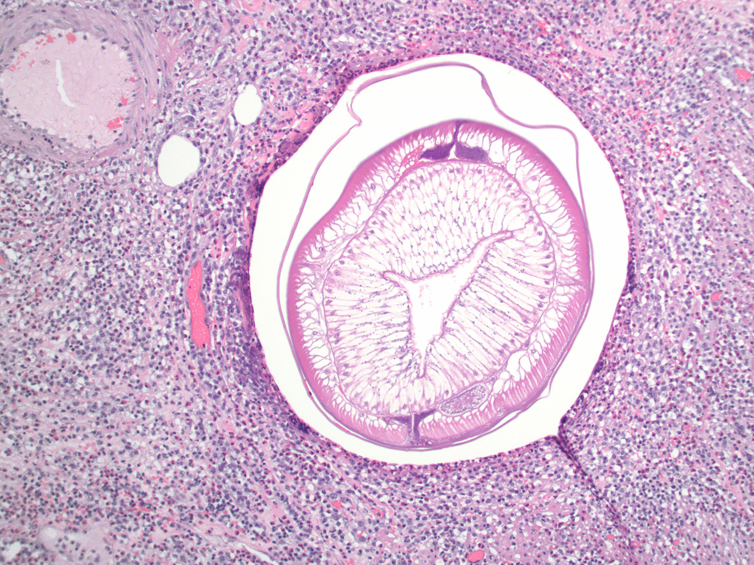


Answer to the Parasite Case of the Week 751: Anisakis species.
Thanks to all who provided comments on this case! Idzi kicked us off with his comment: "The Y-shaped lateral chords tell me that this is patient has eaten raw (or undercooked) fish! 😉". Florida Fan then elaborated, "The cross sections tell us that this is a nematode. From online publications and Idzi's comment, the very tall musculature and Y-shaped lateral chords favor a diagnosis of anisakiasis."
Here is an annotated image showing some of the key features:

This week's case is an interesting challenge! It was donated by Drs. David Sullivan and Ted Nash. The patient is a young girl from Ethiopia with intermittent GI distress. Her parents said that she had passed several ovoid objects per rectum. One of these objects was submitted for histologic sectioning and revealed the following.


Answer to the Parasite Case of the Week 750: Bertiella sp. proglottids and egg.
Kudos to everyone who got this right! As noted by Idzi, "At first I thought this would be plant material, but the egg is quite typical for Bertiella sp. showing the typical pyriform apparatus inside. Secondly, in the third histology picture I think I see calcareous corpuscles, typical for and consistent with a cestode structure, together with an eosinophilic acellular tegument. So I'll follow Florida Fan with the diagnosis of Bertiella sp. infection.
Here is the characteristic egg with the pyriform apparatus inside:

Our previous case (565) shows this in greater detail, along with the short and wide proglottids (with a disturbing resemblance to bow tie pasta).
When looking at the proglottids from cases 565, one can see how sectioning may reveal the 'interesting' histopathologic appearance that we see in this case.
Previous case:


