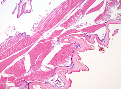
A 56-year old male who had just returned hom from a safari in Uganda presented with a red swollen nodule over the right iliac crest and a 1.5 cm x 0.5 cm white-tan object was removed and submitted to surgical pathology. The following are H&E stained sections of this object:
20 times original magnification

20 times original magnification

40 times original magnification

100 times original magnification

100 times original magnification

100 times original magnification
Identification?


8 comments:
Just got back?
This should be myiasis: Tumbu fly (Cordylobia anthropophaga)larva. This is a clinical diagnosis and generally doesn't require surgical biopsy. Occlusion of "breathing hole" with vaseline and tweezers to remove. Tungiasis is in the differential, but unlikely here.
Hi Mundele Djo,
Good point - he just got back, but had been there for 2 weeks if that helps.
Pictures number one and number four clearly show the mandibular parts of the larva. This case is therefore a myasis. Thanks to Mundele Djo, we now learn about another causative agent. Funny that the "man eating bug" also devours animals.
Florida Fan
You can see the hooklets on the body of the larva, which are positioned randomly over it's body.
It would have been obvious that thus would be Cordylobia when examined under a stereomicroscope.
Please give these species to right people ! (so not pathology :) )
So pathology!
Try to find someone to ID something odd like this - starting now you have 5 minutes, IDing source will have to pay for shipping, 3rd party billing is a must, and have you paid up malpractice if it was melanoma...
This is a huge problem for the people several layers closer to the patient -- I know of no easy catalogues of sources to advise with some of these many weirdnesses. Public Health is lame and even for straightforward malaria is days. CDC has a website, typically is responsive, but is not near real time and very time consuming.
So as a simpleminded pathologist I can make a section for near nothing, get paid maybe $30 or at least not be out of pocket, get to "some kind of bug dx", and if is a complicated/interesting case or some complications try to track down a source.
Parasite Gal would be a great source as obviously will take on anything but is not in many Rolodexes and even though there is a well known lab in Rochester there are lots of process issues.
Yes this is slightly a rant but really a rant of frustration that it is not easier to find easy to use algorithms and sources to help ID odd things like this.
Richard Garcia-Kennedy MD
Dear Dr. Garcia-Kennedy,
I understand completely! A lot of the ability to ID these critters is to have a good quality microbiology lab available. Barring that, you are left with a variety of options which can include tradition Surg path sectioning and time consuming send-outs. Fortunately, the CDC DPDx team now offers e-consults with a 24 hour turn around time or less. They're a great group and can work from photos of histologic sections or the intact specimen. I highly recommend them!
Post a Comment