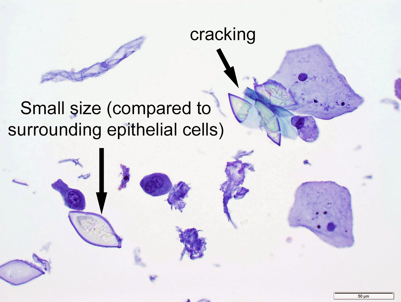
This was a challenging case given that uric acid crystals are a very convincing mimic of Schistosoma hematobium eggs. Fortunately they can be differentiated by a number of features:
- The crystals are pointed at both ends while S. hematobium eggs have a single (terminal) spine.
- The pointed ends of the crystals are more triangular, while true S. hematobium spines have a pinched-off appearance.
- Crystals are smaller than Schistosoma hematobium eggs (usually < 100 micrometers in length; compare their size to the surrounding epithelial cells) and vary significantly in size. In contrast, S. hematobium eggs are large (112 - 170 x 40-70 micrometers).
- There are no internal staining structures on Papanicolaou stain, unlike what would be seen with true eggs (see example below).
- Crystals are often cracked, whereas true eggs rarely crack but instead may be crushed or deformed
- Crystals are very birefringent on polarized light unlike true eggs which do not polarize to this extent.
Comparison of true S. hematobium egg (left) and crystal (right) on Papanicolaou stain:
Uric acid crystals may be seen in urine in a number of settings, particularly in acidic urine and certain metabolic states such as gout.
From Blaine Mathison:
These ellipsoid-shaped
structures might look like schisto
but really they're uric acid
crystals, dont ya know
high concentrations in blood
might lead to gout
so from your diet leave excess
sugars out
or excruciating pain you may
feel down in your toe!


+and+crystal+(right).jpg)


No comments:
Post a Comment