Answer to the Parasite Case of the Week 675: Coenurosis, a tapeworm infection with the larval form of coenurus-forming Taenia species, such as T. multiceps (the most commonly implicated species), T. serialis, T. brauni, and T. glomeratus.
Some of you noted the resemblance of this tissue infection with cysticercosis. That makes sense since human cysticercosis is due to the related cestode, Taenia solium. The primary difference is the presence of a single protoscolex in cysticercosis, vs. multiple protoscoleces in coenurosis.
Here is my approach to the diagnosis:
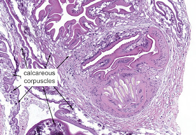
- We can first tell this is a cestode by the presence of the calcareous corpuscles (nice call Idzi!)
- The presence of a thin eosinophilic tegument and loose stroma is also supportive of this being a cestode.
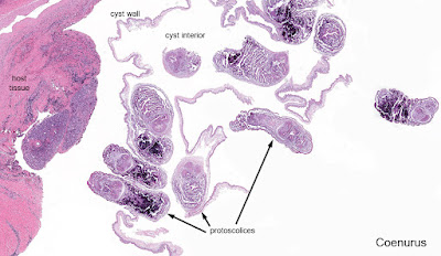
- We can further narrow this down to a larval cestode based on its location (within tissue) and cystic appearance. The cystic appearance helps us further narrow down our differential to cystic echinococcosis, cysticercosis, and coenurosis.
- The presence of multiple protoscoleces allows us to rule out cysticercosis (which only has a single protoscolex), bringing us to a differential diagnosis of just 2 cestode infections, cystic echinococcosis and coenurosis.
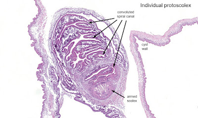
- Finally, we can differentiate cystic echinococcus from coenurosis by the appearance of their protoscoleces. Taenia spp. protoscoleces have a convoluted spiral canal, whereas Echinococcus spp. do not.


3 comments:
Love the detailed breakdown on the differential !!! Clear, Concise. Coherent. Much Appreciation and Many Thanks! Cheers!
Thank you Dr. Pritt for your very educational detailed explanation. We learned a lot from you.
Florida Fan
You're very welcome Sean and Florida Fan! Thank you for being part of our awesome parasitology community!
Bobbi
Post a Comment