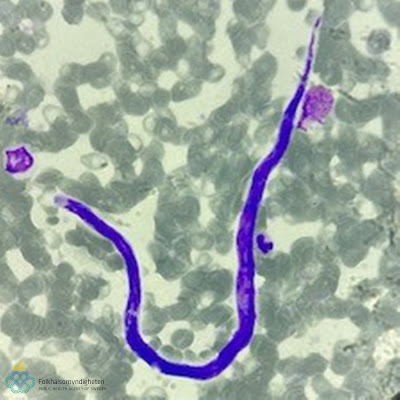
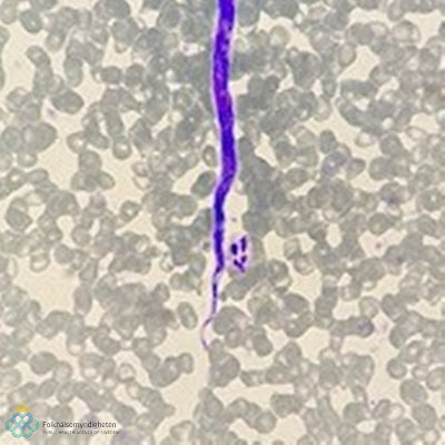
This week's case is generously donated by Dr. Ioana Bujila of the Public Health Agency of Sweden. The patient is a 67 year old woman from Gabon. Blood was examined by direct mount and Giemsa-stained blood films, and the following were identified:
These objects are approximately 228 micrometers in length.
What is your diagnosis? Are there any additional laboratory analyses that are recommended in this case?


11 comments:
Lymphatic Filariasis
Microfilaria of Loa loa or Mansonnella perstans.
Microfilaremia is the main factor that drives the endemicity level in a given area. It can be used as a surrogate for transmission assessment before and during mass drug administration. If microfilariae are found in the peripheral blood, fertile adults must be viable and drug distribution must continue if transmission is to be interrupted.
PCR can be used to identify species.
Loa loa microfilaria. Co-infection with Onchocerca volvulus needs to be considered before initiating treatment.
From the images and videos, I think it could be Loa loa microfilariae. The microfilariae of this species are sheathed, something that can be seen in the second video. In Giemsa-stained smears, however, the sheath is not stained, which is quite common in this stain (except for Brugia malayi). The geographical area of origin is also compatible with this species.
Perhaps LAMP could be used to confirm identification.
Interesting case as always, with very good photos and videos.
Luis H.
Watching the live, second video one can clearly see the head of the microfilarium move inside a sheath. This rules out the non sheathed microfilaria like Mansonella or Onchocerca or Drancunculus. We only have to choose between the sheathed Brugia, Wucheria and Loa loa. For sure the two Brugias are geographically too far away to be considered. The tail in the Giemsa smear shows the column of nuclei extending all the way to its end, this eliminates any chance for it to be a Wucheria. The only identification left to us is Loa loa. This is such a not commonly encountered organism that we have to appreciate its educational value
Florida Fan
Microfilariae of Loa loa.
Loa loa
It’s sheathed microfilaria- brugiya malayi, loa loa or wuchereria bancrofti are three options.
The nuclei seems to present till the tip of the tail. This rules out brugiya which has 2 distinct nuclei separated by a thin delicate thread like filament, and Wuchereia which has well defined nuclei that does not occupy the tail. This most likely is Loa loa microfilaria
I’d categorize this one under “wonderful” 😀
(I wish I scienced harder to know what this is)
These are by far the most beautiful images I have ever seen from what is definitely Loa loa microfilariae. I especially LOVE the video (second one) where the sheath is nicely visible!
Step-by-step identification is nicely explained by Florida Fan. Nothing to add in that regard. I'm pretty sure Bobbi will tell us about the nuclei that "fLowa-flowa" to the tip of the tail ;-)
I may add that a Knott's concentration could be envisaged to obtain a quantitative result before treatment is started. Also a hematoxylin-based staining (like Carrazzi's hematoxylin staining) could be used to demonstrate the sheath more nicely (as Luis H. points out: Giemsa does not usually/reliably stain the sheath of microfilariae) and better show the positioning of the nuclei in the tail.
Superb case!
It appears to have a sheath, and nuclei in a linear fashion at the tail-so maybe Brugia malayi over loa loa?
Post a Comment